Two physicists at the University of Virginia have combined MR and gamma-ray imaging to potentially create a new way of producing high-resolution medical images, according to a study published in Nature.
Called polarized nuclear imaging (PNI), the method is designed to use highly detectable nuclear tracers with the spectral sensitivity and diagnostic power of MRI techniques (Nature, September 29, 2016, Vol. 537:7622, pp. 652-655).
Co-creators Wilson Miller, PhD, and Gordon Cates, PhD, said they have demonstrated the feasibility of the new technique by producing a proof-of-principle image. Rather than imaging protons in water, as with conventional MRI, the pair imaged a radioactive isotope of xenon that has been polarized using laser techniques.
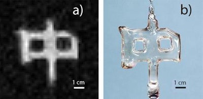
 At left is an image of a sealed glass cell created by University of Virginia physicists by combining MR techniques with the detection of gamma rays emitted by an isotope of xenon contained within the cell. At right is an ordinary photograph of the glass cell. Images courtesy of the University of Virginia.
Once polarized nuclear imaging is refined, the researchers believe it could become an inexpensive way to visualize gas space of the lungs by inhalation of a gas containing the isotopes and the use of PNI to produce an image. The method might also be successful in imaging targeted areas of the body by injecting isotopes into the bloodstream.
Because such small quantities of tracer material would be used, the radioactivity would pose little to no danger to people, the researchers added.
As an example, had Cates and Miller filled their imaging subject -- in this case, a small glass cell shaped like the Chinese symbol for the word "middle" -- with water rather than the radioactive isotope, they would have needed about 10 billion times more water molecules than the number of isotope atoms they used to achieve the same image quality.
This means that with minute quantities of material, they can achieve detailed imagery using MR-based techniques that would otherwise be impossible using a radioactive tracer.