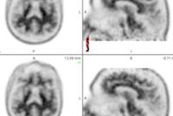
Researchers from Arizona have developed a florbetapir-PET brain imaging technique to better detect the progression of beta-amyloid plaque deposition, which has been linked with Alzheimer's disease.
The findings, published online March 5 in the Journal of Nuclear Medicine, could also help scientists assess antiamyloid treatments being used in clinical trials.
Lead author Kewei Chen, PhD, from Banner Alzheimer's Institute (BAI) in Phoenix, and colleagues analyzed baseline and 24-month follow-up florbetapir-PET scans of 332 people who participated in the Alzheimer's Disease Neuroimaging Initiative (ADNI). The group included normal older adults, as well as those diagnosed with mild cognitive impairment or probable Alzheimer's dementia.
The researchers compared the uptake of florbetapir in the cerebral cortex to its uptake in the cerebral white matter, pons, and cerebellum. Standardized uptake value ratios (SUVRs) were calculated to determine which of the three reference regions offered the best way of tracking amyloid over two years.
The cerebral white-matter reference region provided more accurate tracking of SUVR among patients with probable Alzheimer's, those with mild cognitive impairment, and in older cognitively normal adults who were found to be amyloid-positive at baseline, according to the researchers.
The cerebral-to-cerebral white matter SUVR increases also correlated with poorer scores on participants' neurocognitive tests; SUVRs from the other reference regions failed to demonstrate this pattern. The cerebral white-matter reference region also resulted in less variability in repeated florbetapir SUVR readings over time in the patients who probably had Alzheimer's disease.
Chen and colleagues added that additional studies are still needed to validate the imaging technique for other amyloid PET ligands.