Using PET, Japanese researchers examined a special kind of neural plasticity that allows for motor function recovery during rehabilitation after brain damage, according to a study published in the January 7 issue of the Journal of Neuroscience.
Yumi Murata, from the National Institute of Advanced Industrial Science and Technology (AIST), and colleagues found that motor functions are "remapped" as brain regions farther from a lesion are recruited during early rehabilitation. Functional connections with regions near the lesion are then strengthened during later parts of rehabilitative treatment (J Neurosci, Vol. 35:1, pp. 84-95).
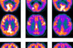
The researchers examined monkeys with injuries to the part of the cerebral cortex that controls hand movements. Murata and colleagues used PET to image regional brain activity in the monkeys as they underwent rehabilitation, scanning them before the injury and at early and late stages of recovery. Activity in the ventral premotor cortex, which is relatively far from the injury, was greater during the early stage of recovery than before the injury, they found.
Additional analysis showed that connections between the lesion and regions of the primary motor cortex immediately surrounding it became stronger in later stages of recovery. The findings could help contribute to the development of new rehabilitation techniques and drugs, as well as new ways to evaluate rehabilitative training, according to Murata.