A new study by Dutch researchers adds evidence that PET imaging with carbon-11-labeled Pittsburgh Compound B (PiB) and FDG can lead to early diagnosis of Alzheimer's disease and differentiate between specific dementias, especially when previous diagnostic certainty is low.
The study, from VU University Medical Center in Amsterdam, found that PiB and FDG-PET improve the clinical diagnosis of Alzheimer's disease patients, and enhance clinicians' confidence in their diagnoses in the setting of the memory clinic.
"The diagnosis of patients in a memory clinic can be complicated, as there can be a lot of overlap in clinical symptoms, particularly in cases of young patients," said lead study author Dr. Rik Ossenkoppele from VU's Department of Neurology and Alzheimer's Center, who presented the findings in June at the Society of Nuclear Medicine and Molecular Imaging annual meeting.
Prominent biomarkers
In recent years, both FDG and PiB have become more prominent biomarkers in the diagnosis of several forms of dementia, Ossenkoppele noted. PiB is designed to detect deposits of amyloid plaque in the brain, which have been associated with the onset of Alzheimer's disease. FDG measures metabolic activity of the brain at rest and has been used to distinguish between several forms of dementia.
"However, FDG and mostly PiB studies to date have been conducted in highly selected diagnostic groups," Ossenkoppele said. "The aim of the present study is to assess the impact of PiB and FDG-PET on the diagnostic process in patients from a memory clinic, encompassing a wide spectrum of cognitive and/or behavioral symptoms."
Researchers enrolled a total of 154 patients with a mean age of 64 years. The first part of the single-center study was designed to establish a clinical consensus diagnosis by using standard dementia screening tools, such as neurological examinations, brain MRI exams, and psychological evaluations.
The study tallied 66 Alzheimer's patients (44 male, 22 female), 30 people with mild cognitive impairment (23 male, seven female), 15 individuals who had experienced subjective memory complaints (seven males, eight females), and 18 subjects with frontotemporal dementia. In addition, there were five patients with Lewy bodies, 10 people with other forms of dementia, six individuals with other psychiatric disorders, and four individuals with neurological diseases.
Diagnostic confidence
Cognitive neurologists used standard dementia screening tools to diagnose patients, and then scored their diagnostic confidence in their interpretations. Ratings ranged from an average of 69% for the group with mild cognitive impairment to 79% for patients with subjective memory complaints. Diagnostic confidence for Alzheimer's patients was 71%.
Researchers then proceeded to the PET scans. First, patients were imaged with PiB for 90 minutes to obtain parametric images of the biomarker binding to potential clusters of amyloid. FDG-PET scans were then conducted 45 to 60 minutes after injection and lasted for 15 minutes. Standardized uptake value (SUV) ratios were calculated throughout the brain.
The resulting PET images were visually assessed by a nuclear medicine physician and reported to the neurologists to determine how the additional information changed their initial clinical diagnoses and their confidence in those interpretations.
As a result of the scans, the neurologists' diagnoses changed in 35 (23%) of 154 patients. "Most changes occurred in the Alzheimer's disease group, as two-thirds remained stable," Ossenkoppele said.
In the Alzheimer's group, PET with PiB or FDG confirmed a total of 44 (67%) of the 66 cases prior to PET imaging. The second-greatest change was among patients with frontotemporal dementia. PET with PiB or FDG confirmed 11 (61%) of the 18 cases prior to PET.

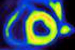
 The image on the left shows PiB binding (red) in the cortical regions of the brain to indicate the presence of amyloid plaque, while the image on the right shows PiB binding to white matter (red), which signifies a negative result. All images courtesy of Dr. Rik Ossenkoppele.
PiB-PET scans were positive in 40 (61%) of 66 patients with clinical Alzheimer's and in five (28%) of 18 patients with clinical frontotemporal dementia. FDG SUV patterns matched the clinical diagnosis in 58% of patients with Alzheimer's and 33% of patients with clinical frontotemporal dementia, the authors noted.
The study also found that diagnostic confidence increased from 71% in initial PET scans to 87% after PiB and FDG-PET imaging.

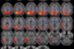
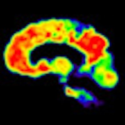
 FDG-PET images show a normal brain (top) with normal functions (red) compared with an abnormal brain (bottom) with a diagnosis of Alzheimer's and lack of FDG uptake (yellow and green).
In addition, two-year clinical follow-up in a subsample of 39 patients showed that PiB and FDG predicted progression to Alzheimer's disease for patients with mild cognitive impairment, and that the diagnosis of dementia established after PET remained unchanged in 96% of the patients.
The researchers found a "frequent mismatch between clinical diagnosis and PET findings," Ossenkoppele said. "PiB and FDG led to a diagnostic change in almost one out of four patients [and] increased the diagnostic confidence, and PiB and FDG seem most useful when diagnostic confidence is low," he concluded.