A Japanese study highlights the benefits of a new PET radiotracer for imaging the proliferation of cancer cells in non-small cell lung cancer (NSCLC), as compared with PET's workhorse radiopharmaceutical, FDG. The new tracer may also help noninvasively reveal the DNA synthesis of NSCLC cells.
The radiotracer 4'-[methyl-11C]-thiothymidine, or carbon-11 4DST (C-11 4DST), was developed by Dr. Jun Toyohara and colleagues from the Positron Medical Center at the Tokyo Metropolitan Institute of Gerontology specifically to image cellular proliferation. Uncontrolled proliferation is a distinctive characteristic of cancer cells.
In a study presented at the recent RSNA 2011 meeting, researchers from the department of radiology at Yokohama City University, led by from Dr. Ryogo Minamimoto, compared C-11 4DST to FDG, which is by far the most widely used PET agent for oncology. The researchers believe that C-11 4DST could give clinicians more detailed information about cell proliferation in individual cancers, enabling them to better direct a patient's therapy.
Study protocol
The researchers enrolled 18 patients: 12 men and six women, with a mean age of 69 years. Each patient received a PET/CT scan (Biograph 16, Siemens Healthcare) with intravenous injection of C-11 4DST (18 mCi to 20 mCi), and a PET/CT scan with intravenous injection of FDG (10 mCi).
Three images were acquired over intervals of five, 20, 40, 60, and 80 minutes after injection with C-11 4DST. Image acquisition at the 40-minute mark was common among all patients. FDG-PET/CT images were obtained 60 minutes after injection.
"FDG, representing glucose metabolism, is an excellent PET tracer and [is] the standard for cancer imaging," said Minamimoto. "However, understanding characteristics of each cancer is now required for selecting appropriate therapy for cancer patients."
PET can provide information about cancer noninvasively, and FDG uptake has a relatively high correlation with cellular proliferation. "So, we set FDG as the standard reference to evaluate the potential of 4DST for representing cellular proliferation," he said.
Minamimoto and colleagues compared maximum standardized uptake value (SUVmax) for C-11 4DST to that of FDG for lung tumors. They compared results from both imaging techniques to measurements of the antibody MIB-1, which detects Ki-67, a cellular marker for proliferation, in pathological specimens after surgery. The group also compared the results with CD31 expression, as a measure of cancer cells.
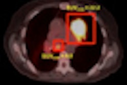
The results showed pathologically proven NSCLC in all 18 patients, with a total of 19 lesions. There were 16 cases of adenocarcinoma, two squamous cell carcinomas, and one large cell cancer.
SUVmax results
The mean SUVmax with C-11 4DST for lung cancer was 2.9 ±1.0 (range, 1.5-4.7), compared with a mean SUVmax for FDG of 6.2 ± 4.5 (range, 0.9-17.3).
The correlation between SUVmax and MIB-1 index was also greater with 4DST (r = 0.67) than with FDG (r = 0.60). In addition, the correlation between SUVmax and the number of cells positive for CD31 was greater with 4DST (r = 0.66) than with FDG (r = 0.59).
The results confirmed a higher correlation with proliferation of lung tumors for C-11 4DST compared to FDG. C-11 4DST PET/CT may also provide noninvasive imaging of DNA synthesis in NSCLC, the researchers concluded.
"The ability to image cellular proliferation would offer the possibility of distinguishing between benign and malignant tissues, measuring tumor activity, and evaluating response to therapy," Minamimoto told AuntMinnie.com. "Moreover, it will predict the prognosis of the cancer patient."
The NSCLC study follows previous research that has found C-11 4DST to be beneficial in imaging other cancers. Toyohara and colleagues concluded earlier this year that the radiotracer was feasible for imaging brain tumors in a small sample of three patients (Journal of Nuclear Medicine, August 1, 2011, Vol. 52:8, pp. 1322-1328).
That study found no serious adverse events in any subjects at any time during the research period. C-11 4DST demonstrated "selective uptake in the bone marrow, which has a high rate of proliferation ... [and] high-level uptake was also seen in the liver," the authors noted. C-11 4DST showed little uptake in normal brain tissues, which resulted in low background activity for imaging brain tumors.