Skeletal imaging with sodium fluoride (NaF) PET provides "superior imaging" compared to standard nuclear imaging techniques and can help physicians accurately investigate the cause of bone pain in children, according to a paper in the September issue of the American Journal of Roentgenology.
An evaluation of NaF-PET scans by researchers from Children's Hospital Boston and Massachusetts General Hospital found that NaF-PET provides an "excellent alternative" to tracers such as technetium-99m (Tc-99m) used for radionuclide bone scintigraphy (AJR, Vol. 197:3, pp. 713-719).
NaF-PET is the modality of choice for bone scans at Children's Hospital, said lead study author Dr. Laura Drubach, assistant professor in the hospital's department of radiology. "Our child abuse cases, for example, are done with sodium fluoride, and many patients are referred for bone pain for sports medicine, such as the evaluation of trauma and stress fractures," she told AuntMinnie.com.
NaF-PET cases
Drubach estimated that more than 500 exams have been conducted at the facility using NaF-PET. Whole-body scans or images of a particular region of interest are acquired in either the 2D or 3D mode (Advance Nxi, GE Healthcare), beginning 30 to 45 minutes after NaF injection. Both emission and transmission scans are used for attenuation correction.
Children's Hospital typically uses a 60-mCi/kg (2.2-MBq/kg) dosage of NaF, with a minimum dose of 300 mCi (11.1 MBq) and a maximum dose of 4 mCi (148 MBq).
The patient population is younger than 25 years old, with teenagers accounting for the majority of individuals with sports-related bone injuries. Child abuse patients at Children's Hospital are predictably much younger.
Drubach and colleagues last year published a study on child abuse and the efficacy of NaF-PET compared to baseline skeletal surveys. The researchers found that NaF-PET produced sensitivity and specificity rates of more than 90% in the overall detection of fractures related to child abuse, when compared with baseline skeletal surveys (Radiology, April 2010, Vol. 255:1, pp. 173-181).
With young patients, because of radiation exposure and ethical issues, it is "impossible to do a head-to-head comparison of technetium-99m [methylene diphosphonate (MDP)] and sodium fluoride on the same patient, since it would give radiation twice," Drubach said. "So, we had to compare what was described in the literature with child abuse cases."
 |
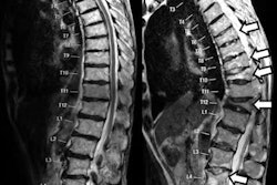
| Images are of a 14-year-old male with low back pain. Maximum intensity projection (MIP) image (A) of the lower thoracic and lumbar spine and pelvis shows increased uptake at the left pars interarticularis (arrow) that is consistent with pars stress fracture. Sagittal slice (B) and transverse slice (C) show the localization of uptake (arrow) in the pars articularis. All images courtesy of Dr. Laura Drubach. |
Injection to imaging
One of the advantages of NaF-PET imaging is the radiopharmaceutical itself, which has "superior characteristics compared to technetium-99m bone scintigraphy for MDP: greater bone concentration -- about twice that of MDP -- and faster clearance from the blood, which allows for earlier imaging than MDP," Drubach said.
Given the quick uptake of NaF, imaging can be performed within 30 to 40 minutes of injection, Drubach said. With Tc-99m MDP, the waiting time to imaging after injection can be as long as two to three hours. The radiopharmaceutical also clears from the blood quickly and is evacuated from the body through the kidneys.
Still, there are precautions that should be taken with the use of NaF in PET scans. "The intrinsic radiation exposure of sodium fluoride -- millicurie by millicurie -- is greater than with technetium, so you do have to be careful with the technique," Drubach added. "In our study, we decreased the dosage of the sodium fluoride, so the radiation exposure is equivalent to standard MDP. You still can get excellent images [with sodium fluoride PET]. In all our cases, radiation exposure was no higher with sodium fluoride than with MDP."
 |
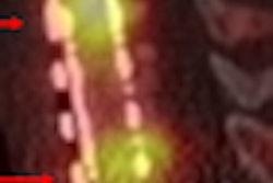
| Images of a 15-year-old male with mid and upper back pain show increased uptake in the spinous process (A) (arrow). The condition is confirmed on the coronal (B), sagittal (C), and transverse (D) images. |
 |
In conclusion, Drubach and colleagues wrote that they believe the "excellent image quality obtained with NaF-PET skeletal imaging makes it an attractive option for the evaluation of suspected bone trauma and stress-related injuries."
While the authors currently have no other studies under way using NaF-PET imaging, Drubach suggested that oncology is one potential area for additional research into the efficacy of the radiopharmaceutical, especially in adult patients.
Last year, the Society of Nuclear Medicine (SNM) published guidelines for performing and interpreting NaF-PET studies, which stated that NaF is useful for detecting skeletal metastases. NaF-PET was also deemed appropriate for a number of benign disease indications in select patients (Segall et al, Journal of Nuclear Medicine, November 1, 2010, Vol. 51:11, pp. 1813-1820).