One of the most prominent figures in nuclear medicine in the last half-century is Dr. Henry Wagner Jr. of Johns Hopkins University in Baltimore, MD. Dr. Wagner has conducted pioneering research in imaging the perfusion and ventilation of the lungs, kidney and spleen scanning, myocardial perfusion scanning with potassium-43, gated blood pool imaging, and imaging brain receptors with PET. His new autobiography, A Personal History of Nuclear Medicine: Reflections of a Pioneer (Springer, New York City, 2006), tracks his many accomplishments and provides his perspectives on the challenges facing the academic and medical establishments.
I chose nuclear medicine to be my medical specialty in 1957. As did all of us who chose to become specialists in nuclear medicine, we encountered many obstacles to achieving our goals. Many of the basic principles of the new medical specialty had not yet achieved widespread acceptance by the medical establishment. Anatomy, radiology, and surgery remained the foundation of medical practice.
I first encountered nuclear medicine when I arrived in London in July 1957, five years after I graduated from Johns Hopkins medical school. Nuclear medicine was not then a recognized medical specialty.
 |
| Dr. Henry N. Wagner Jr. has been responsible for a number of major advances in the course of his history in nuclear medicine. |
The general public had heard the term "atomic medicine," which was linked in the mind of the public with the development of the atomic bomb during World War II. The medical use of radioactive materials was based on the same scientific principles that produced the atomic bomb, and there was an underlying fear by the public of anything that had to do with radiation.
These negative perceptions lingered long after the end of the war. It would be decades before nuclear medicine would find its place in medical practice and biomedical research, before nuclear medicine would be able to define itself as a scientific discipline. When the medical profession eventually understood what the specialty was really all about, nuclear medicine had moved medicine beyond its dominant focus on anatomy to the new focus on "molecular medicine."
More than any other specialty, nuclear medicine brought together structure and function, which accounts for the wide acceptance today of combined structural, functional, and biochemical imaging in medicine and biomedical research. As Arthur Koestler has written: "In biology, what we call structures are slow processes of long duration; what we call functions are fast processes of short duration." They are all processes, that is, changes in mass as a function of time.
The story of the birth and growth of nuclear medicine is one of the most fascinating in medicine, an example of the observation that things don't happen; people make things happen. Nuclear medicine evolved using the tools of physics and chemistry to solve patient problems. But first, political, as well as scientific and technological, challenges had to be faced and solved.
The most fundamental principle of nuclear medicine, the "tracer" principle, was invented in 1913 by a Hungarian chemist, Georg von Hevesy. He discovered that it was possible to "track" molecules as they participate in chemical processes within the living body. The idea is that molecules can be made to emit "photons," analogous to radio signals, as they move around within the body, telling us what they are doing as they take part in chemical reactions essential for all life.
Imaging thoughts
On July 14, 1978, our team at Johns Hopkins submitted a grant proposal to the National Institutes of Health entitled "Multi-User Application for Specialized Research Equipment," in order to obtain funds to purchase a cyclotron (Cyclotron Corporation Mode CP-16) for our nuclear medicine division.
All proposed projects were directed toward in vivo imaging of neuroreceptors, starting with C-11 spiroperidol and C-11 diprenorphine. We would concentrate on Parkinson's disease, Huntington's disease, tardive dyskinesia, schizophrenia, and chronic pain.
In our grant proposal we stated, "We believe that the ability to study noninvasively such neuroreceptors in man will lead to fundamental new information of unique importance.... Although the concept that mental function is basically chemical is not new, recent technical developments have made it possible for the first time to attempt to track the miniscule amounts of chemicals within the brain and map out where and how chemical reactions are occurring in the living brain."
On February 8, 1979, Dr. Bob Heyssel, director of Johns Hopkins Hospital (formerly head of nuclear medicine at Vanderbilt University), wrote to me:
"I happened to see the article in the (Baltimore) Sun in which you are quoted extensively concerning expansion of your brain study program for thought imaging.... Don't we have enough problems as it is without our faculty and others helping to make us look like idiots or incompetents or worse?"
Bob had read an article written by Jon Franklin, a Pulitzer Prize-winning reporter from the Baltimore Sun, in which he had written: "Johns Hopkins scientists say they will install a scanner capable of watching the chemical process of thought as it flickers through the brain.... It is designed to help map the circuitry of thought and allow scientists to understand how information is processed by the brain.... Once brain specialists understand the normal flow of energy through the brain, they should be able to pinpoint the problem in patients with certain diseases, including schizophrenia, senility, Huntington's disease, Parkinson's disease, manic-depressive psychosis, drug addiction and manganese poisoning....
"Dr. Henry Wagner, the scientist in charge of the project, expects the device, a positron emission tomography (PET) scanner, to pinpoint chemical malfunctions in the brain and, for the first time, to allow scientists to tinker directly and rationally with the chemical engines of thought.... 'If we find something that runs too fast, we can treat it by slowing it down. If it's too slow, we can speed it up.'"
On May 25, 1983, four years after I received Bob's letter, we succeeded in carrying out the first successful imaging of a neuroreceptor in the living human brain. Using the radioactive tracer carbon-11 N-methyl spiperone (C-11 NMSP) and PET, we were able to image the accumulation of a radioactive tracer by dopamine receptors in the basal ganglia of my brain. The chemical synthesis of the tracer and the PET imaging were carried out in the Nuclear Medicine Division at Johns Hopkins Hospital.
Human guinea pig
Carfentanil is the most potent of all opioid drugs and is potentially toxic or even lethal to human beings. It is recommended that any game animal which has been given the drug for immobilization should not be consumed for at least 45 days. Some state that it is probably not wise under any circumstances to consume animals that have been immobilized with carfentanil, and there have been reports of humans developing symptoms of drug toxicity from consuming animals given immobilizing drugs. As little as 20 μg (micrograms, an almost invisible drop) of carfentanil is lethal to human beings.
We knew carfentanil was an extremely potent opiate because of the experience of its use in immobilizing wild animals. It had never been given to a human being. We thought (correctly, it turned out) that it would be avidly bound by the subject's opiate receptors.
We had carried out the first imaging of a neuroreceptor (the dopamine receptor) in a human being (I was the subject then, too) in 1983. The carfentanil imaging study of my brain was performed in May 1984. It was the first imaging of an opiate receptor in a human being.
We had carried out prior studies in rodents and baboons prior to the first human study. The dose of carfentanil that I was to be given would be exceedingly small, but we thought it wise to have an anesthesia machine present in case there was an effect on my breathing. As it turned out, there was some constriction of my pupils, but no other effects, so we scheduled the PET imaging procedure for the next day in the nuclear medicine department.
As I lay with my head held in position in the NeuroEcat PET scanner, I was injected with 25 millicuries of C-11 carfentanil, containing 80 nanograms/kg of the drug. In the room were Jim Frost, Sol Snyder, Jon Links, our chief nuclear medicine technologist Jim Langan (who had been in nuclear medicine at Hopkins since its inception in 1958), and two other technologists, Jay Rhine and Martin Stumpf.
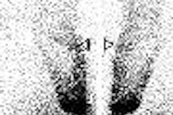
Three imaging planes were viewed, with the cephalic plane passing through the cerebral cortex above the corpus callosum; the central plane passed through the caudate nucleus, putamen, thalamus, and cerebral cortex; and the lower plane passed through the cerebellum. Images were obtained for an hour after injection of the tracer.
After 30 minutes, the greatest accumulation of radioactivity was in the medial thalamus, with less in the lateral thalamus. The frontal and temporoparietal cerebral cortex had somewhat less activity, but substantially more than the occipital cortex. In the caudate slice, there was activity in the pituitary and temporal lobes of the cerebral cortex.
Three hours later, naloxone was injected at a dose of 1 mg/kg, followed by a second dose of 25 millicuries of C-11 carfentanil. The radioactivity accumulation was markedly decreased compared to that in the earlier study before naloxone. This provided strong evidence that the carfentanil was binding to mu opiate receptors.
A healthy skepticism
Under the peer-review system, individual faculty members can bypass the power structures of medical schools, and there has been a steady, annual increase in the NIH budget, which led to a great increase in the size of medical school faculties and increasing numbers of research projects. The steady increase in the number of creative basic and clinical scientists throughout the United States resulted in the U.S. achieving the major role in biomedical research.
Individual creativity was the hallmark of biomedical research. Essential to the success of the peer-review system is the expertise and honesty of the reviewers, the objectivity and confidentiality of the proposals, and the objective opinions of the peers. Their reviews were returned to the investigators, and resubmissions were commonplace if the initial application failed to be funded. Scientists worked not to gain the favor of their academic or political leaders, but to gain the respect and approval of their peers from throughout the country.
We young investigators soon learned the importance of having convincing evidence that our ideas would work by obtaining and submitting preliminary results. Otherwise the proposal could be rejected if one of the reviewers stated that the basic premise was unfounded. Usually a young person beginning to carry out research would join the team of an experienced, well-funded investigator. Only later would he or she strive to become a principle investigator.
Today, with increasing clinical demands for the faculty in medical schools, it is difficult for clinical faculty to have time to do research. They are now required to generate income for the institution by providing patient care, and are reduced to "spending a day in the lab."
More and more research grants are being awarded to those with both an M.D. and a Ph.D., or with a Ph.D. only. Some clinicians, who are basically not effective in carrying out research, do so only to be able to climb the ladder of academic medicine. Sadly, after they become professors, their research activities often come to an end.
Fortunately, I had seen at an early age the excitement and pleasure of research. I had the good fortune to work in the laboratory of the famous psychobiologist Curt Richter as a student, and then join the stimulating surroundings of the NIH from 1955-1957. My being a medical student and then a "house officer" in internal medicine at Hopkins also encouraged a love and respect for research, which Johns Hopkins has always valued as a principal function of the university and its medical school. No other environment could have better prepared me for a clinical research career.
As clinical associates at the Heart Institute, we were encouraged to conduct research projects, both on our own and under the leadership of our superiors, while caring for the patients of other investigators. I was surprised to observe that some research scientists hesitated to have contact with patients, to the point of conducting "rounds" of patients without entering the patient's room.
One of my first lessons at the NIH was that the opposite of "to love" is not "to hate," but "to ignore." When we presented our work to our peers and leaders, their critical comments were an indication that they took the work seriously.
Once, after a colleague had presented some research results, the research director of the Heart Institute at the time made no comments. On the way out of the conference room, I asked: "Do you agree with that?" I was disappointed when he told me that he didn't agree with any of what had been said, but chose not to comment. I learned then that one's best friends are those who will tell you when you are wrong, as well as when you are right.
While on the house staff at Hopkins, I had always taken the words of our superiors as "gospel." I was disabused of this attitude during my stay at the NIH. Even today, I still tend to believe that what people tell me is not true. Not that they want to deceive me, but that they "don't know what they don't know."
This skeptical attitude is helpful to a professional scientist. When my students or fellows were to make a scientific presentation or lecture, I encouraged my students and colleagues to ask: Is what I am saying clear? Is it true? Is it new? Is it significant?
By Dr. Henry N. Wagner Jr.
AuntMinnie.com contributing writer
June 4, 2006
Copyright © 2006 AuntMinnie.com