Spatial maps of the brain captured on functional near-infrared spectroscopy (fNIRS) revealed increased brain activity in students performing surgery after virtual training, according to a study presented at the American College of Surgeons (ACS) annual meeting in San Diego last month.
Researchers from Rensselaer Polytechnic Institute (RPI), Harvard University, and the University at Buffalo used the noninvasive imaging capability of fNIRS to create spatial maps of the brains of medical students as they performed a surgical procedure on cadavers.
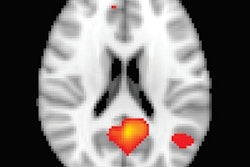
 Functional near-infrared spectroscopy brain images of medical students while performing surgery after no training (control), training with a physical simulator (FLS), and training with a virtual simulator (VBLAST). Image courtesy of Arun Nemani.
Among the 19 medical students evaluated, five received no training, six practiced on a physical simulator, and eight practiced on a virtual simulator. Brain imaging showed that the medical students with training had much higher activity in the primary motor cortex when performing surgery than the students without training.
"This is a significant leap in the use of noninvasive brain imaging technology to quantify human motor skills and represents a paradigm shift in which surgeons and other medical professionals may be certified and credentialed one day," co-author Suvranu De, PhD, said in a statement from RPI.
What's more, students with virtual training completed tasks in an average of 13.05 minutes, which was significantly faster than the 15.5-minute average for the group without training. Students with physical training completed the surgery faster still -- averaging 7.9 minutes.
"These results demonstrate that optical neuroimaging provides quantitative and standardized metrics for assessing surgical skills acquisition and expertise," co-author Xavier Intes, PhD, said. "Optical neuroimaging is uniquely positioned to play a central role in developing tailored surgical training programs for optimal skill acquisition and retention assessment."