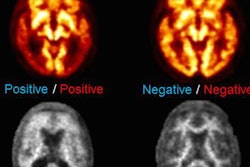
PET scans can track the stages of Alzheimer's disease in cognitively normal adults, according to a new study published in the journal Neuron.
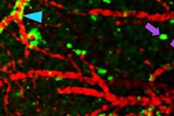
The study findings also shed light on the nature of tau and amyloid protein deposits in the aging brain. The accumulation of beta-amyloid plaques has long been considered the primary culprit in Alzheimer's disease, but over the past decade, tau, a microtubule protein important in maintaining the structure of neurons, has emerged as a major factor, lead investigator Dr. William Jagust, of the University of California, Berkeley, and colleagues wrote (Neuron, March 2, 2016).
Jagust's group conducted PET scans of 53 adults. Five of these were between the ages of 20 and 26, 33 were between the ages of 64 and 90, and 15 were patients ages 53 to 77 who had been diagnosed with probable Alzheimer's dementia.
The results of the scans were consistent with the neuropathological stages established by German researchers Heiko and Eva Braak through postmortem analysis of the brains of suspected Alzheimer's patients. These stages range from 1 to 6 and describe the degree of tau protein accumulation in the brain.
The PET scans included in the study confirmed that with advancing age the Alzheimer's-linked tau protein accumulated in the medial temporal lobe, which is home to the hippocampus and the memory center of the brain. The study showed that higher levels of tau in this area were associated with greater declines in episodic memory.
The study findings indicate that tau imaging could be an important tool in helping to develop therapeutic approaches that target the correct protein depending on the disease stage, the team concluded.