Dual-time-point FDG-PET/CT is not an effective way to differentiate between benign and malignant salivary gland tumors, according to a study from Japan in the September issue of the American Journal of Roentgenology.
The analysis showed that benign salivary gland tumors had high FDG uptake, similar to that of malignant salivary gland tumors, wrote lead author Dr. Akira Toriihara, from the department of diagnostic radiology and oncology at Tokyo Medical and Dental University, and colleagues.
Limited benefit?
FDG-PET/CT is the accepted standard when it comes to evaluating malignant tumors in the head and neck; however, it can have limited benefit when distinguishing between benign and malignant salivary gland tumors, according to the authors.
And while some research has found that dual-time-point FDG-PET/CT is useful in differentiating benign from malignant head and neck lesions, other papers have discouraged its use for characterizing thyroid nodules.
"To the best of our knowledge, there have been no published reports of evaluation of the usefulness of dual-time-point FDG-PET/CT for salivary gland tumors," Toriihara and colleagues wrote (AJR, September 2013, Vol. 201:3, pp. 639-644).
Imaging protocol
The researchers retrospectively evaluated 21 women and 18 men (mean age, 51.6 years) with salivary gland tumors who underwent FDG-PET/CT at the hospital between August 2007 and July 2011. They assessed a total of 40 salivary gland tumors (one patient had two tumors).
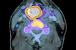
 Images are of a 38-year-old woman with a left submandibular gland tumor. A maximum intensity projection image (A) and fused PET/CT image (B) were obtained. Early-phase SUVmax, delayed-phase SUVmax, and retention index for the tumor were 17.4, 23.1, and 32.8%, respectively. The tumor was diagnosed as pleomorphic adenoma. Images courtesy of AJR.
The patients fasted for at least four hours before receiving an injection of 3.7 MBq/kg of FDG. The early-phase set of PET/CT images (Aquiduo, Toshiba Medical Systems) of the head and neck from skull base to subclavicular area were acquired 60 minutes after injection. The scan protocol was repeated for delayed-phase imaging 120 minutes after FDG injection.
Early-phase maximum standardized uptake value (SUVmax), delayed-phase SUVmax, and retention index were compared for benign and malignant tumors. The retention index of FDG uptake in each tumor was calculated as follows: [(delayed-phase SUVmax - early-phase SUVmax) / early phase SUVmax] × 100%.
Correlations between the delayed-phase SUVmax and retention index in benign, malignant, and all tumors were analyzed, and differences with p-values less than 0.05 were considered statistically significant. In addition, the study group used receiver operator characteristics (ROC) analysis to determine the diagnostic accuracy for malignant salivary gland tumors.
SUVmax comparisons
In their analysis, Torriihara and colleagues found some benign tumors that expressed very high FDG uptake, and they eventually concluded there were no significant differences between the benign and malignant tumors in mean early-phase SUVmax or mean delayed-phase SUVmax. The mean retention index of the malignant tumors, however, was significantly higher than the index for benign tumors.
| SUVmax for malignant vs. benign tumors by time point | ||
| Benign tumors | Malignant tumors | |
| Mean early-phase SUVmax | 7.3 (± 4.4) | 9.2 (± 4.5) |
| Mean delayed-phase SUVmax | 8.3 (± 5.8) | 11.2 (± 5.9) |
| Mean retention index | 8.5% (± 12.3%) | 20.1% (± 10.2%) |
The ROC analysis found no significant difference in diagnostic accuracy between the delayed-phase SUVmax and retention index. When a cutoff value of 15% was used for the retention index, sensitivity, specificity, and accuracy each were 75%. The authors wrote that those levels "were not satisfactory for clinical decision."
"In conclusion, dual-time-point FDG-PET/CT is not useful for discriminating between benign and malignant salivary gland tumors, because benign tumors also show high FDG uptake, which increases during the delayed phase," Torriihara and colleagues wrote.