Assessment of heart muscle viability using PET/MRI yielded comparable results to PET/CT in a study presented this week at the Society of Nuclear Medicine and Molecular Imaging (SNMMI) annual meeting.
"[The research] showed that MR can be used for PET attenuation correction in the same way that CT can," said lead author Dr. Jeffrey M.C. Lau, PhD, from Washington University in St. Louis, in a statement.
The highly sensitive soft-tissue contrast of MRI and the functional and metabolic imaging of PET are particularly valuable for cardiac indications and could pave the way for the hybrid modality's use in a variety of applications.
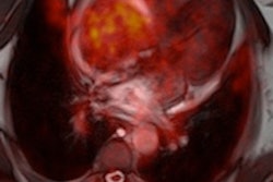
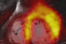
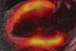
In the current study, the researchers compared the visualization of FDG in cardiac cells with PET/MRI compared to PET/CT.
Lau and colleagues analyzed 31 patients with no history of heart problems, who were screened for cancer. All subjects underwent both PET/CT and PET/MR with FDG administered about an hour before PET/CT and two hours before PET/MRI.
FDG uptake in the myocardium was measured by looking at a cross section of the left ventricle of the heart. The average measurement of FDG uptake in the left ventricle was nearly identical for PET/MR (4.68) and PET/CT (4.62).
Additional studies are needed to collect more information about the benefits and appropriateness of PET/MRI in clinical practice, Lau and colleagues noted.
This study was conducted in conjunction with Siemens Healthcare.