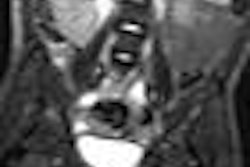
Is your cardiac MRI scanner a $2 million patient gurney? It might be if you're not effectively using every available moment to prepare and scan patients, eliminating downtime between exams, according to a presentation at the recent Society for Cardiovascular Magnetic Resonance (SCMR) meeting.
When it comes to throughput, many physicians and MRI technologists always want to get more done in less time, noted Dr. Scott Flamm, head of the section of cardiovascular imaging in the Cleveland Clinic radiology division. At the same time, that goal is tied to obtaining the best diagnostic information for patient care.
"MR has a sort of albatross around its neck in that we are able to do everything, but we don't need to do everything," Flamm said during his presentation at the San Francisco meeting. "From a bottom-line perspective, if we really want to do this quickly and efficiently, we have to remember we are focusing on the clinical question. We need to answer the clinical question."
Prescan suggestions
Much of the time saving can be done even before the MRI exam begins. To ensure that patients arrive early or on time for their appointment, call them the day before and detail how they should prepare for the scan, especially if sedation is required, Flamm recommended.
The list of prescan requirements should include making sure a patient does not have any caffeine-based products, such as chocolate, coffee, or tea, if a stress test will be performed. Also, the patient should fill out a safety questionnaire in the waiting area, rather than in the MRI scan room.
MRI personnel should calculate the patient's estimated glomerular filtration rate (eGFR) well before the patient is on the scanner. In addition, the insertion of any intravenous lines should be handled in the nurses' area.
Flamm said placing IV lines is a very simple step in the prescan process, but one that is often overlooked.
"While other processes are going on in the background from the previous scan, we are using our MRI table to put an IV line in a patient," he said, making the MRI scanner a "$2 million gurney."
Wired or wireless electrocardiograph chest leads also should be placed on the patient before he or she enters the MRI room.
Technology optimization
The use of acceleration techniques such as parallel imaging cannot be emphasized enough, according to Flamm. Practices that use these tools work faster "and considering how good the technology is today, we have very few limitations and very few downsides in using the technology," he said.
The key is to include acceleration techniques in imaging protocols to acquire more images per time period, rather than acquiring higher-resolution scans in the same amount of time. There isn't always a need for higher spatial resolution in MRI, but the acquisition of more information in the same amount of time will provide answers to the patient's clinical issue much faster.
One key imaging technique is to acquire multiple image slices with each breath-hold. For example, a 1.5-tesla MRI scanner using acceleration techniques can acquire two to four slices per breath-hold. On a 3-tesla scanner, four to six slices per breath-hold can be achieved.
"On a 3-tesla scanner, that means two breath-holds to acquire all the cine sequences we need," Flamm said. "That makes a considerable difference and decrease in the amount of time."
There are also "acceptable compromises" in that "incredibly fine spatial resolution" is not always needed to solve the clinical question, he added. High spatial resolution "is great for research studies, and in some situations it is valuable, but for most general clinical purposes, we do not need high spatial resolution or even extremely high temporal resolution."
By optimizing imaging protocols, MRI scans can become more efficient in determining a specific clinical outcome for a patient's disease or condition. At the Cleveland Clinic, physicians use standardized protocols for specific indications, which are referred to as "exam cards."
"We have a preset series of sequences, orientations, and angulations that we go through each and every time for that particular suspected disease," Flamm said. "We only make minor modifications for each pulse sequence that comes up."
He also advised that physicians limit research sequences in an MRI scan for the sake of time and expediency.
"If what you are doing is part of a grant or a specific research project, then it would make sense [to add research sequences]," he said. "But if you are trying to set up an efficient system, you can pull these [sequences] out to answer the specific clinical question."
Fill the downtime
Patient imaging time can also be reduced by eliminating as much downtime as possible, Flamm said. Every single moment between patient appointments should be filled with productive activities by MRI personnel, such as prepping the next scan by changing linens and reloading contrast agent.
To avoid repeat MRI scans, Flamm recommended positioning a qualified technologist at the scanner in case an imaging sequence needs to be repeated or if a different approach is required during the scan to avoid mistakes or inadequate results.
"To be able to do this quickly and efficiently, we need to have an adequate number of highly skilled technologists who can scan with consistent efficiency," Flamm added.
It is also very important to monitor performance. Each quarter the Cleveland Clinic evaluates how long it takes, on average, to perform each diagnostic MRI study.
With each procedure, "we need to think about how we can shave every little second off the process," he concluded.