Computer-aided detection (CAD) software can outperform both a senior thoracic radiologist and a resident trained in chest imaging for detecting nonsolid pulmonary nodules larger than 3 mm, according to research from Pitié-Salpêtrière Hospital in Paris.
The results support the use of a CAD system as a second observer for both nonsolid and solid nodules detected on CT studies, according to Dr. Catherine Beigelman-Aubry.
Nonsolid nodules, which may represent either non-neoplastic or neoplastic lesions, have a higher prevalence of malignancy than solid nodules; ground-glass opacity (GGO) nodules represent approximately 20% of the total nodules demonstrated in screening programs, Beigelman-Aubry said.
To evaluate CAD software for detecting these nonsolid pulmonary nodules, the researchers reviewed 12 CT exams of 10 patients, each of whom had at least one solid nodule. Seven of the studies were performed on a LightSpeed 16-slice CT scanner (GE Healthcare, Chalfont St. Giles, U.K.) and reconstructed with a lung kernel and slice-thickness interval of 1.25 mm/0.6 mm.
Five studies were performed on a Brilliance 40-slice CT scanner (Philips Healthcare, Andover, MA) and reconstructed with a 0.8-mm/0.4-mm lung kernel and slice thickness, Beigelman-Aubry said. She presented the research at the 2007 RSNA meeting in Chicago.
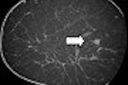
Two experienced thoracic radiologists working in consensus identified 13 solid nodules and 265 nonsolid nodules. The researchers elected to focus their research on 95 of the nonsolid lesions that ranged in size from 3 to 29 mm, because the LMS-Lung CAD software (Median Technologies, Sophia Antipolis, France) utilized in the study was not optimized to detect nodules smaller than 3 mm, according to Beigleman-Aubry.
Of these 95 nodules larger than 3 mm, 63 were GGO nodules and 31 were part-solid. The 12 CT scans were first read by a senior chest radiologist and a resident trained in chest imaging in a blinded study. The scans were then subsequently analyzed by CAD.
The senior chest radiologist identified 49 (52%) of the nonsolid nodules, while the resident identified 42 (45%). CAD identified 58 (61%). When the CAD results were added to the findings of the readers, the percentage of correctly identified lesions climbed to 82% and 79%, respectively.
"There were only five false-positive markings," Beigelman-Aubry said. "Four of these corresponded to large, ill-defined areas of GGO. The fifth corresponded to a band of GGO. Overall, the positive predictive value of the CAD in detection of nonsolid nodules was 95%."
As the research only included a limited number of patients, the results need to be confirmed in a larger study, Beigelman-Aubry said.
By Cynthia Keen
AuntMinnie.com contributing writer
February 27, 2008
Related Reading
Hybrid lung segmentation software boosts performance, February 13, 2008
Experience may make a difference with lung CAD, February 4, 2008
CAD offers value in detecting lung nodules with CT, December 17, 2007
CAD may boost radiologists' ability to characterize lung nodules, November 25, 2007
CAD performs well in lung nodule detection, February 5, 2007
Copyright © 2008 AuntMinnie.com


