Multiple myeloma, a treatable but incurable form of bone marrow cancer, is slowly giving way to new drugs that are extending and improving the quality of patients' lives, according to a report from the 2007 Congress of the European Hematology Association (EHA) in Vienna.
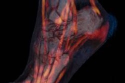
Better treatment options make accurate evaluation all the more important for patient management. And according to a new study, multidetector-row CT (MDCT) detects multiple myelomas slightly better than MRI, and significantly better than PET. Neither MRI nor MDCT can do the job alone, however.
New drugs
"Twenty years ago only 40% of our patients responded to our therapy," Professor Heinz Ludwig from Vienna's Wilhelminenspital said in a June 8 statement. "Now we have a range of drugs that are effective individually or in combination in more than 90% of the people treated. Patients who could no longer be helped by any other means were able to get back out of bed with these new drugs and lead a normal life."
Ludwig is the hospital's director of oncology and hematology, and president of the 2007 EHA congress.
Multiple myeloma is so named because its foci often form concurrently at several places in the skeleton, Ludwig explained. The initial steps of malignant transformation occur in B lymphocytes, the precursors of plasma cells, reducing their usefulness as an intelligent component of immune system.
Two therapeutic agents in particular have shown promise, and more than 80 new drugs are in the development pipeline, Ludwig said. First, the active ingredient bortezomib kills malignant plasma cells by inhibiting the degeneration of important regulatory proteins in cancer cells, while promoting the rebuilding of bony tissue. The second agent, lenalidomid, is an immunomodulating substance that inhibits inflammation, slows the spread of degenerated cells, and is anti-angiogenic.
New imaging results
Dr. Jin Hur and his colleagues from Yonsei University College of Medicine in Seoul, South Korea, sought to compare the efficacy of MDCT with MRI and FDG-PET in 10 patients with stage III multiple myeloma.
"Computed tomography is a well-established but rarely used technique for imaging multiple myeloma," Hur and his colleagues wrote (Journal of Computer Assisted Tomography, May/June 2007, Vol. 31:3, pp. 342-347). Most studies have been conducted with single-detector scanners, but MDCT and the ability to create 3D MPR images offer great potential for evaluating the extent of osteolytic bone lesions, they added.
The researchers retrospectively evaluated 140 vertebrae in 10 patients (7 men, 3 women; ages 50-77, mean age 62) with newly diagnosed multiple myeloma, all of whom underwent MDCT and MRI of the spine and FDG-PET exams within two weeks' time. All were diagnosed as stage III based on marrow biopsy and staged according to Durie-Salmon criteria.
Noncontrast CT was performed on a Somatom Sensation 16 scanner (Siemens Medical Solutions, Erlangen, Germany) at 16 x 0.75 collimation and 5-mm reconstruction increments with 5-mm section widths, 120 kVp, and 200 mAs. For image reconstruction, a smooth medium convolution filter was used.
MRI images were acquired on a Siemens Magnetom Vision 1.5-tesla scanner equipped with a surface coil. Sequences included T1-weighted spin echo (repetition time/echo time of 600/12 msec, 512 x 80 matrix, 3-mm slice thickness). T2-weighted spin-echo MRI was also performed (repetition time/echo time 3900/112 msec, 1024 x 330 matrix, 3-mm slice thickness), according to the authors.
FDG-PET studies were performed on an Allegro scanner (Philips Medical Systems, Andover, MA) following a minimum six-hour fast and injection of 370 MBq of F-18 FDG, the researchers wrote. They acquired whole-body scans 40-60 minutes after injection of the agent using multiple positions. An iterative transmission algorithm was used to reconstruct 3D attenuation-corrected transaxial images using a 3D image filter and 128 x 128 image matrix.
Two experienced radiologists reviewed the MDCT and MRI images independently, resolving assessment differences by consensus, the group stated. At MDCT the researchers looked for punched-out lytic lesions, expansile lesions with soft-tissue masses, and diffuse osteopenia, using the classification established by Laroche et al and modified by Mahnken et al. At MRI the radiologists looked for focal, multifocal, or diffuse areas of decreased signal intensity of the bone marrow relative to adjacent normal tissues on T1-weighted images, and areas of increased signal of the bone marrow on T2 images. Three nuclear medicine physicians read the FDG-PET images, looking for diffuse or focal areas of increased FDG uptake.
Respectively, MDCT, MRI and PET detected the following numbers of lesions: 102 of 1040 vertebrae, 95 of 140 vertebrae, 84 of 100 vertebrae. "The difference between the abilities of MDCT and MRI to detect lesions was not statistically significant (p = 0.289)," the authors wrote, noting a statistically significant difference between MDCT and FDG-PET (p < 0.001). MDCT was also superior to MRI for detecting small osteolytic lesions 5 mm and smaller (p = 0.031).
MRI and CT are complementary modalities for multiple myeloma, the authors wrote. Radiography continues to play an important role in Durie-Salmon staging for newly diagnosed patients, but it is ineffective in detecting early disease, often underestimating bone involvement, particularly in the spine.
Durie-Salmon PLUS incorporates advanced imaging modalities, a positive development in that detection of a single additional lesion can change a patient's disease stage. In previous studies, Mahnken et al found that MDCT allowed a better evaluation of areas at risk of fracture than MRI, while Horger et al concluded that whole-body MDCT was appropriate for diagnosing lytic bone changes and assessment of fracture risk.
"In our study, MDCT was very sensitive in detecting small osteolytic lesions in the spine as compared with MRI and FDG-PET," the group wrote. "However, evaluation of osteopenia by CT is nonspecific and might lead to a misinterpretation of the imaging findings if MDCT is used without a correlation to MRI."
For its part, whole-body FDG-PET exams can potentially detect medullary and extramedullary lesions in a single exam; however, PET is costly and requires CT or MRI anatomic correlation, they added.
"Although MRI is still the imaging modality of choice for the evaluation of myelomatous spinal lesions, it can underestimate bone marrow involvement of the spine in multiple myeloma, especially for small osteolytic lesions, which is critical for accurate staging and proper management," Hur and colleagues concluded.
By Eric Barnes
AuntMinnie.com staff writer
June 18, 2007
Related Reading
No survival benefit of high-dose therapy in multiple myeloma, January 5, 2006
MRI sees spinal metastases earlier, but low-dose whole-body MDCT aces skeletal workup, May 20, 2005
MDCT staging may obviate bone scintigraphy in some patients, March 6, 2005
Single-fraction radiotherapy effectively treats painful bone metastases, October 20, 2003
Single-fraction radiotherapy the treatment of choice for painful bone metastases, February 11, 2003
Copyright © 2007 AuntMinnie.com