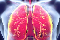
Lost in the enthusiasm over CT lung screening's mortality benefit was a big question: how to handle follow-up. With false-positive rates over 25% in some screening studies, there has to be a better way to separate patients who may have cancer from those who don't. In a July 2 article in PLoS One, researchers think they may have found one.
In a study of nearly 1,200 at-risk individuals screened with CT at New York University over nine years, researchers found that increased age, smoking history, and evidence of emphysema as indicated on pulmonary function tests are the best ways to determine which patients with suspicious nodules should be followed up and which ones shouldn't. The results could help guide future lung screening programs.
"The challenge ... is to develop a screening strategy that will identify those at highest risk of lung cancer, detect small lung nodules in these individuals, specify whether the nodule is malignant, and distinguish those malignant nodules that will progress from those that may be more indolent and could be managed more conservatively," wrote a group led by Dr. Alissa Greenberg (PLoS One, July 2, 2012).
20% mortality benefit
While the finding in the National Lung Screening Trial (NLST) of a 20% mortality benefit for CT screening of high-risk smokers has been much touted, the study also produced a 24% detection rate for noncalcified nodules, of which 96% were found to be false-positive results. Better profiles for who should be screened with CT -- as well as which nodules should be followed up and which ones simply monitored -- would go a long way toward reducing both the cost and anxiety of CT lung screening by reducing the number of follow-up CT scans, the group wrote.
Greenberg led a research group from the university's Lung Cancer Biomarker Center that sought to identify the risk factors that might predict which patients would have nodules on CT screening, and help clinicians determine which of these nodules might represent an early lung cancer. For a study population, they included 1,182 individuals who received CT lung screening between March 2001 and June 2010. All participants were older than 50 years (mean, 63 years) and had more than 20 pack-years of smoking history (mean, 42 pack-years); some also had asbestos exposure due to referrals from a local utility workers union.
Individuals received low-dose CT screening exams, initially on four-detector-row scanners (Somatom Volume Zoom, Siemens Healthcare) and later on 16- and 64-row scanners. The researchers employed a low-dose technique with 40 to 80 mAs and 7-mm-thick sections reconstructed at 6 mm; additional high-resolution 1-mm sections were acquired for suspected lung nodules.
The patients also performed forced expiratory spirometry tests to measure lung function, and they completed questionnaires that included questions on demographic characteristics, tobacco use, occupational exposure, alcohol use, and family history, among other factors. If no noncalcified nodules were detected on a screening exam, individuals were invited to annual screening studies for the first three years, then every two years thereafter.
Follow-up of noncalcified nodules suspicious for cancer was based on nodule size, initially according to guidelines proposed by the Early Lung Cancer Action Program (ELCAP), and later based on Fleischner Society guidelines:
- Individuals with nodules larger than 8 mm were referred for evaluation, including possible PET scans and biopsy.
- Participants with 6- to 8-mm nodules received a follow-up CT scan at three to six months, another scan at nine to 12 months, and a final scan at 24 months if no changes were seen.
- Those with 4- to 6-mm nodules received follow-up CT at six to 12 months and again at 18 to 24 months.
Nodules were considered benign if they were stable for more than two years, and individuals were invited to continue annual or biennial screening. Subsolid nodules larger than 6 mm were never considered benign, and if they were stable after two years, continued CT follow-up every two years was recommended. Radiologists reviewing the CT screening exams were also asked to note the presence of other abnormalities, such as emphysema or signs of asbestos exposure.
For the first arm of the study -- identifying risk factors that would indicate the presence of nodules on initial screening CT -- the researchers compared individuals with noncalcified nodules larger than 4 mm to those who had no nodules. They found that increasing age, male gender, and history of emphysema were the only statistically significant predictors of the presence of solid and subsolid nodules.
The second part of the analysis focused on those patients with noncalcified nodules that turned out to be actual cancer; Greenberg and colleagues compared them to individuals with benign nodules to identify the risk factors that might indicate malignancy. The researchers diagnosed 33 lung cancers in 30 patients, with the control group consisting of 128 subjects who had nodules that were presumed to be benign. Subsolid nodules were excluded from this analysis because previous research has linked these with malignancy.
The group found that reduced lung function as measured by forced expiratory volume (FEV1) as well as forced vital capacity (FVC) on spirometry tests was a statistically significant predictor of malignancy. Lung cancer patients scored 89.9% and benign individuals 96.5% (p = 0.0129) on FVC tests; for FEV1 tests, the scores were 84.3% for malignant cases and 93.9% for benign cases (p = 0.0068).
The number of pack-years of smoking was another statistically significant predictor, with the lung cancer cases reporting a mean of 56.5 pack-years, while the benign cases reported 40.0 pack-years (p < 0.0001). The individuals with malignant disease also had more and larger nodules. Current smoking status, presence of emphysema, and the ratio of FEV1 toFVC were statistically insignificant predictors.
In a secondary finding, the screening period used in the NLST -- three years of annual CT screenings in high-risk individuals -- might be too short, according to the authors, as all but one of the incident cancers diagnosed in the current study were found after more than three years of normal CT scans.
The authors recommend that their findings be used to help guide work up of small noncalcified nodules found on CT screening. These individuals should be assessed for lung function, smoking history, nodule type, and size and number of nodules.
"This data can be used to begin to refine the ideal population for screening," they wrote. "Age, smoking history, and presence of emphysema should all be considered when deciding whether to recommend lung cancer screening."