Conebeam CT (CBCT) surpassed MDCT for diagnosing hearing loss related to superior semicircular canal dehiscence (SSCD), a small hole in the bony wall of the inner ear bone that can cause dizziness and hearing loss, researchers reported at the American Roentgen Ray Society (ARRS) meeting on Wednesday.
The Belgian study included 21 patients who underwent both conebeam CT (Newtom 5G, Inline Systems) and MDCT (Discovery CT750 HD, GE Healthcare) of the right and left temporal bones, according to Dr. David Volders, from AZ Sint-Jan Brugge, and colleagues.
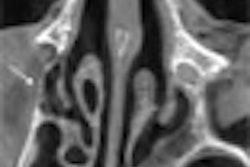
The study compared conebeam CT, which has a spatial resolution of 0.125 mm, to MDCT, with a spatial resolution of 0.23 mm, for detecting 28 anatomic structures, otosclerosis, and SSCD in the temporal bone.
Conebeam CT scored better for the visualization of every anatomic structure in the axial and coronal plane of the temporal bone, with the most significant differences in the coronal plane, the group reported. CBCT scored significantly better than MDCT in visualizing normal temporal bone anatomy.
CBCT corrected a false-negative diagnosis for otosclerosis in 17.9% (5/28) of cases and a false-positive diagnosis for SSCD in 25% (7/28). The average CT dose index volume (CTDIvol) for an examination with CBCT was 26.84 mGy, compared with 56.05 mGy for MDCT.
Conebeam CT provides superior detail in the study of temporal bone anatomy, is more sensitive than MDCT for otosclerosis, and offers higher specificity for the detection of SSCD, Volders and colleagues concluded. In addition, the radiation dose of CBCT was less than half that of MDCT, making conebeam CT the exam of choice, they noted.