Severe H1N1 pneumonia has a distinct imaging signature, according to University of California, Los Angeles researchers, who presented their work at the Society of Critical Care Medicine meeting earlier this month in Houston.
In the largest analysis to date of severe H1N1 pneumonia requiring intensive care, the team found that all 18 patients had abnormal radiographic and CT findings. In addition, 100% of the patients had ground-glass opacities and consolidation on CT on admission to intensive care units (ICUs).
Dr. Nader Kamangar, a pulmonary and critical care specialist at Olive View-UCLA Medical Center; Dr. Cecilia Jude, a thoracic radiologist; and several fellows and residents conducted the retrospective study of patients during the 2009 H1N1 pandemic.
"Compared to prior studies of H1N1 patients requiring intensive care, this study demonstrated a higher rate of ground-glass opacities and greater involvement of middle and lower lung zones," Jude noted.
All 18 individuals were admitted to the ICU with a primary diagnosis of H1N1. Their ages ranged from 23 to 62 years (mean 41). Each had chest x-ray on admission, and five also had a CT scan.
The diagnosis was confirmed in each patient with either a rapid influenza detection test or a real-time reverse transcriptase polymerase chain reaction assay. Twelve (67%) of the patients met the criteria for acute respiratory distress syndrome and required mechanical ventilation. Three (25%) of these individuals died.
On chest x-ray, 16 (89%) of the patients had ground-glass opacities, 16 (89%) had consolidation, and six (33%) had reticular opacities. Seventeen (94%) patients had bilateral radiographic abnormalities.
The abnormalities involved primarily the middle (78%) and lower (100%) lung zones. Sixteen (89%) had extensive chest x-ray abnormalities involving three or more lung zones. The team also found pleural effusions in 16 (89%) patients.
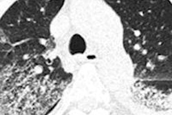
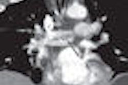
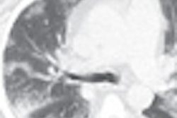
The most common CT findings were consolidation and ground-glass opacities, seen in all patients. All the CT abnormalities were bilateral and extensive, involving three or more lung zones.