Apple's iPad has the hardware power to support preliminary interpretation of emergency brain CT studies, but an inability of current software to access patient information and previous imaging can lead to some missed findings, according to researchers in Ireland.
Investigators from Cork University Hospital found 93% diagnostic agreement between general radiologists performing preliminary interpretation of brain CT scans on an iPad and neuroradiologists providing final reads on a conventional 2-megapixel monochrome LCD. Discrepancies were attributed primarily due to the lack of access to clinical information and previous images on the iPad.
"Our results are encouraging and indicate that from a hardware perspective, the iPad display is suitable for preliminary emergency department CT brain interpretation," said Dr. Patrick McLaughlin.
He presented the research during a scientific session at the European Congress of Radiology (ECR), held earlier this month in Vienna.
While Apple's iPhone has become an invaluable asset to many radiologists, and initial trials have shown merit for mobile image interpretation, radiologists at Cork University Hospital do not use the device for clinical image interpretation, McLaughlin said. This is largely due to its small display size and lower spatial resolution.
Because the American College of Radiology (ACR) recommends that a device's display resolution should at least equal the number of pixels in the image of interest, the iPhone's resolution of 420 x 380 pixels has approximately 40% fewer pixels than required to display a CT image at its native resolution of 512 x 512 pixels, McLaughlin said.
The iPad, however, provides resolution of 1024 x 768, which is five times more than the number of pixels in the iPhone, and its physical size is almost three times larger, according to McLaughlin.
"Many radiologists and application developers quickly identified that the Apple iPad may be a favorable candidate for mobile image review," he said. "There are already numerous applications available."
However, simply accepting the iPad display on the basis of its specifications alone may not adequately reflect its efficacy in radiological image interpretation, McLaughlin said. As a result, the researchers sought to create a study design to better demonstrate the iPad's clinical efficacy.
In the first phase, the study group evaluated the iPad by semiobjectively quantifying its display performance using a contrast-detail phantom. The researchers selected Artinis CDRAD 2.0 phantom, which has been found to determine precisely a display's threshold for low-contrast detectability and detailed resolution, he said.
One point is awarded for correctly identifying the location of each lucent dot. Although the iPad had the lowest score of 141 points when image magnification was disabled, enabling zooming capability increased its score by 16% to 163 points. That score was not a statistically significant difference from the 165 points produced on the diagnostic display.
Subjectively, the iPad's touchscreen zoom function was judged to be very user friendly. "The tactile nature of interaction allowed quick and reliable navigation through the test pattern," McLaughlin said.
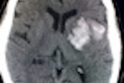
In the second part of the study, the researchers compared the diagnostic efficacy of the iPad with a diagnostic monochrome display in 100 consecutive emergency brain CT studies referred from the institution's emergency department. Of the 100 studies, 77% were normal.
All studies were acquired on a GE Healthcare LightSpeed VCT 64-slice CT scanner without intravenous contrast.
Brain CT studies were selected primarily because of the ongoing requirements for timely preliminary CT analysis and to triage patients for further advanced imaging and intervention, McLaughlin said. Blinded to imaging request details and previous imaging, two general radiologists performed preliminary interpretation in consensus using the iPad and Mobile OsiriX software.
Final interpretation was then performed by one of two neuroradiologists using a Barco Coronis 2-megapixel diagnostic monochrome display. The neuroradiologists had full access to the imaging request details and prior imaging.
All interpretative discrepancies were recorded and scored using the ACR's RadPeer scoring system, McLaughlin said. Image transmission to the iPad took approximately seven seconds over the institution's wireless network, and no technical issues were experienced.
The researchers found total diagnostic agreement between interpretations on the iPad and 2-megapixel display in 93% of cases. Of the seven imaging findings that were missed on the iPad, four were deemed to be understandable misses that were not clinically significant.
"These included three subcutaneous hematomas and a one periventricular white-matter hypodensity," he said. "Importantly, these abnormalities were subsequently identified following a second read with the clinical information available."
Two other misses -- an acute right lentiform nucleus ischemia and a congenital craniocervical fusion -- were understandable but also clinically significant, McLaughlin said. However, subsequent review of the images with access to the patient's previous studies led to identification of the abnormality.
In general, the researchers found that the iPad did not impede the radiologists' workflow. Interpretation time was not delayed by the loading and unloading of images and no technical issues were experienced.
Discrepancies were predominantly attributed to the lack of clinical information and previous imaging available to the radiologist at the time of interpretation, McLaughlin said.
"We look toward the software developers and look forward to further testing and other applications and updates to the current applications which will resolve these issues," he concluded.