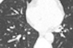
By analyzing CT values in the affected lung regions, quantitative CT (QCT) is improving the diagnosis of chronic obstructive pulmonary disease (COPD) to enable physicians to tailor treatment to individual patients, according to a study presented at the 2010 RSNA meeting in Chicago.
Colorado researchers found that QCT is strongly associated with the degree of spirometric impairment in cigarette smokers -- particularly the degree of air trapping.
The mortality, morbidity, and treatment costs are rising for COPD, which some experts predict will take its place as the third leading cause of death within a decade. COPD involves at least two distinct disease processes: a small-airway disease component in the form of inflammation that can be seen indirectly at CT, and a second component in the form of parenchymal destruction characterized by loss of alveolar attachments and reduced elastic recall that is also visible on CT as emphysema.
The contribution of these two processes varies widely from patient to patient, and reliable methods of pinpointing the disease type in specific lung regions have been lacking. QCT has shown the potential to quantify distinct components of COPD -- particularly emphysema and air trapping, said Joyce Schroeder, MD, from National Jewish Health in Denver in her RSNA presentation.
"These [components] may be important for phenotyping the disease and subsequently devising individualized treatment," she said. "Our goal is to come up with a CT phenotype in the COPD gene study, and we're going to correlate them with physiologic measurements of airway obstruction."
COPD is defined at spirometry as FEV1/FVC < 0.7. In this equation, FEV1 (forced expiratory volume in one second) represents the amount of air that can be forcibly exhaled in the first second of a forced expiration. FVC (forced vital capacity) represents the amount of air that can be forcibly exhaled after taking the deepest breath possible. Within these results, the GOLD scale (Global Initiative for Chronic Obstructive Lung Disease) is used to stratify COPD severity from mild COPD (GOLD1) to very severe COPD (GOLD4).
"We start with DICOM images at 16 bits per pixel, and we extract features that are important in terms of the radiological disease that we're looking at and the parameters clinicians are interested in," Schroeder explained.
The study data were taken from the COPDGene Study, a large multicenter trial that will perform genome-wide analysis to identify specific genotypes associated with the phenotypes of COPD determined by quantitative imaging, she said. More than 10,000 patients have been enrolled in the study, including 6,000 patients with COPD, stratified by severity, and 100 normal controls.
To date, more than 9,150 CT scans have been received. From these data, Schroeder and her team analyzed 2,273 datasets, including 1,001 patients with GOLD0, 213 patients with GOLD1, 531 patients with GOLD2, 340 patients with GOLD3, and 188 patients with GOLD4.
From the CT data, feature extraction was used to define the portion of the lungs affected by emphysema and air trapping, as follows:
- Emphysema = %lung ≤ 950 Hounsfield units (HU) on inspiratory CT
- Air trapping = %lung ≤ 856 HU on expiratory CT
The subjects underwent volumetric CT at full inspiration (120 kVp, 200 mAs) and again at the end of a normal expiration (120 kVp, 50 mAs), using various multidetector-row CT scanners.
QCT analysis including lung segmentation and lobe analysis performed using commercially available software (VIDA Diagnostics, Iowa City, IA). Schroeder and colleagues also performed a multivariate analysis to evaluate the relationship between demographic, clinical, and QCT variables and FEV1 percent predicted and FEV1/FVC ratio.
Analysis based on the whole lung revealed that air trapping was more prevalent than emphysema. The relative (r2) values for FEV1/FVC ratio were 0.55 (% emphysema) and 0.71 (% air trapping) p < 0.0001 for all regression slopes, she said. "We noticed that the r2 values are higher for percent air trapping compared to percent emphysema for both spirometry parameters," Schroeder said. "We also see that r2 values are higher for FEV1/FVC ratio compared to the predicted FEV1 percentage."
Analysis by lung lobe revealed higher emphysema measurements in the upper lobes versus air trapping -- and that air trapping was higher in patients who also had higher portions of their lungs affected by emphysema.
"A bivariate [curve] fit of emphysema versus air trapping shows increasing air trapping with increasing emphysema for all five lobes," Schroeder said.
The multivariate analysis of lung lobes found that the FEV1 spirometry measurement was most influenced by lower lobe air trapping and emphysema, while the FEV1/FVC ratio was most influenced by the air trapping component, Schroeder said. These two models accounted for 65% and 74% of the variability in FEV1 % predicted and the FEV1/FVC ratio, respectively.
At the multivariate analysis, significant predictors of reduced FEV1 percent predicted and FEV1/FVC ratio were percent air trapping, BMI, male gender, age at enrollment, and hours of supplemental oxygen use per day.
|
Percent air trapping was the single most powerful predictor of reduced lung capacity based on its proportion of the total variance, according to Schroeder.
Quantitative CT is strongly associated with spirometric impairment in cigarette smokers, she said. "In particular, air trapping on expiration combined with clinical parameters correlates with physiologic meaurement of airway obstruction. On lung lobe analysis, lower lobe emphysema and air trapping are physiologically important," she noted.
FEV1 is most influenced by lower lobe trapping and lower lobe emphysema, while the FEV1/FVC ratio is most influenced by lower lobe air trapping, Schroeder concluded.
By Eric Barnes
AuntMinnie.com staff writer
December 28, 2010
Related Reading
New CAD for COPD parses disease type, severity, October 1, 2010
CAD detects signs of emphysema on DR images, September 27, 2010
CT method measures airway calcium to diagnose lung cancer, June 28, 2010
Altered blood flow in lungs predicts emphysema, April 6, 2010
Imaging shows that even mild COPD reduces cardiac efficiency, January 20, 2010
Copyright © 2010 AuntMinnie.com