SAN FRANCISCO - Among the more important technical innovations to hit multidetector-row CT of late are wide-area detectors. They come with inherent inefficiencies, but they're powering advances in heart, brain, and pediatric imaging that would not otherwise be feasible.
On opening day of this year's International Symposium on Multidetector-Row CT, Dr. Mathias Prokop, chair of radiology at University Medical Center Utrecht in the Netherlands, discussed the implications of wider detectors being produced by CT manufacturers, as well as other technologies that can achieve similar results.
Since 1985, CT performance has been doubling about every two years as measured in anatomic coverage per second. The main reasons are improvements in gantry rotation speed, the advent of dual-source imaging in 2006, and detector width, Prokop said.
"One has to keep in mind, however, that there is a difference between the total width of a detector and the actual width of the detector that you are using," he said. "On a four-slice scanner the [utilized] width would be in the range of 20 mm, but for high spatial resolution you only use 4 mm," he said. All 64-detector-row scanners use the entire scanner width, he said.
 |
| Development of wide-area detectors by manufacturers since 1995, with width in centimeters. The blue lines depict detector width, and the red lines are the widths actually used in imaging applications. All charts and images courtesy of Dr. Mathias Prokop. |
Pluses and minuses
Most 64-detector-row scanners are equipped with 4-cm-wide detectors, but two manufacturers have increased that to 8 cm (Philips Healthcare, Andover, MA) and 16 cm (Toshiba America Medical Systems, Tustin, CA), respectively.
The wider coverage per gantry rotation brings faster acquisition, and the opportunity to do multiple acquisitions at the same location in dynamic scanning, Prokop said. But a disadvantage is the larger anatomic area exposed to radiation for each image.
"There is also potentially more scatter as you widen the detector, and another effect is that the field-of-view determines the z-axis coverage -- something new with these very wide detectors" that requires some planning, Prokop said.
The detectors were developed because "we wanted to cover the heart more rapidly, and [combined with] prospective step-and-shoot techniques reduce the dose substantially to the patient," he said.
While a 4-cm detector can cover the heart in four steps, the 8-cm in two steps, and the 16-cm detector in one step, even a scanner with 16-cm coverage requires two-beat acquisition for faster hearts, meaning retrospective reconstruction and, consequently, a higher dose for many patients.
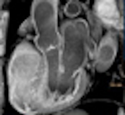
 |
| Single-beat cardiac CT angiography acquisition on a 320-detector-row scanner with 16-cm-wide detectors. |
These days even step-and-shoot image acquisition isn't limited to the heart.
"Single-shot imaging can also be extended to other areas of the body," Prokop said. "It is great for small children -- for example, imaging the thorax takes less than half a second -- and it can also be interesting for trauma patients to image the head, the neck and joints, and for dynamic scans if you want to stay at one spot."
In the brain, a 4-cm detector can cover the basal ganglia in a single rotation, an 8-cm detector the circle of Willis to the corpus callosum, and a 16-cm detector the whole brain.
The shuttle
Wider areas can also be covered using a shuttle mode, which has been implemented by two manufacturers (Siemens Healthcare, Andover, MA, and GE Healthcare, Chalfont St. Giles, U.K.). Anatomic coverage is no longer limited because the scanner table moves back and forth to cover as large an area as needed.
On the other hand, patient movement easily leads to artifact, there is a variable time lag between slices and volumes to accommodate the table movement, he said.
Wider coverage through faster scanning
Another way to achieve wide coverage quickly is to perform ultrafast or flash scanning, either with the second-generation dual-source scanner (Definition Flash, Siemens Healthcare, Malvern, PA) (43 cm/sec) or a single-source scanner that operates at high speeds with a wide-area detector (Brilliance iCT, Philips) (20 cm/sec). Flash scanning is not only helpful for triggered spiral scanning of the heart, as the speed stops fidgety children in their tracks, it also aids trauma imaging in patients for whom every minute is critical. In the case of dual-source scanning, all this is achieved with a detector width of approximately 2 cm.
Reduced geometric efficiency
All helical CT scanners require an extra rotation at the beginning and the end of the scan volume, known as overranging. And while the extra radiation this produces is relatively insignificant for narrower detectors, it becomes a bigger problem with wide-area detectors, Prokop said.
"If you have a wide detector and relatively short areas you want to cover, then the relative dose increase is quite substantial," he said. "The solution to that is dynamic collimators that open and close as you go in and out of range" -- a feature that all of the manufacturers now have that solves the problem neatly, he said.
Wide-area scanners require conebeam reconstruction, which requires the use of smaller z-field-of-views (FOV) or diamond-shaped reconstruction of the z-FOV. As a result, less of the coverage area is reconstructed and more radiation is wasted.
 |
| Geometric efficiency is reduced with the use of wide-area detectors, which require the use of conebeam collimation. Overranging and scatter are increased. |
"That's basically the price that you pay for [wide] area detectors," he said. But the smaller field-of-view works great for children.
Dynamic scanning with wide detectors allows optimal imaging of the heart, joints, and many different kinds of brain protocols, Prokop said.
"It's an absolute advantage, for brain and tumors you can also use the shuttle mode," he said. "Motion assessment in joints is something that is unique to these scanners."
 |
| Clinical applications best suited for use with wide-area detectors, and the recommended detector widths. |
The fast acquisition times are ideal for single and dual-shot heart imaging, and for imaging children and trauma patients. The overranging and scatter phenomena require correction with dynamic collimators and a 2D scatter grid. But the z-FOV is reduced, especially in the periphery of the scan field.
"Step-and-shoot imaging is the main reason why these scanners have been developed, and while there are many emerging applications, the advantages come at a price," Prokop said.
Overall, he added, the total package of features in a scanner is probably more important than the wide-area detectors themselves.
By Eric Barnes
AuntMinnie.com staff writer
May 20, 2009
Related Reading
SCCT publishes cardiac CTA guidelines, May 7, 2009
Fourth-generation MDCT scanners do cardiac differently, July 8, 2008
320-slice coronary CTA permits much lower contrast dose, March 6, 2009
Tube current modulation cuts triple rule-out CTA dose, April 15, 2009
Copyright © 2009 AuntMinnie.com