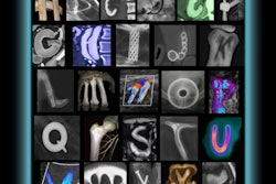
Radiology and art might seem unlikely bedfellows to those in either profession. But the two worlds easily overlap for one artist/medical student, who finds beauty in colorized CT scans of everyday objects ranging from computers to fast food to toy submarines.
Third-year medical student Satre Stuelke started the Radiology Art project in 2007 and has created approximately 30 images over the past two years. The images are displayed on the project's Web site, www.radiologyart.com.
"Basically, the main point of the site is to bring people together through making art with radiological tools," Stuelke told AuntMinnie.com. "The site will hopefully serve as a place where people can share radiological art, and get ideas for more images."
The subjects currently featured on the site fall into three main categories: toys, food, and electronics. "I try [to] pick things that have something beneath the surface that CT can reveal to us, and at the same time, can be meaningful," Stuelke said. He also noted that "the primary goal for all my work is just to make gorgeous images that are fun to look at and think about."
For the toys, it can be their relationship or kinship to human anatomy, Stuelke explained. "Even a windup car has 'organs' that make it go, and has its own functional anatomy which can be easily discovered and examined with the CT scanner," he said.
|
|
| CT scan of a windup rabbit. Video and all images courtesy of Satre Stuelke and radiologyart.com. |
| Having trouble viewing this clip? Click here to download the free Flash player. |
The images of personal electronics on the site include an Apple iBook, an iPhone, and a Palm PDA, and Stuelke noted that he likes scanning electronics to which we perhaps become overly attached.
"Some people just can't live without their iPhone. When it all comes down to it, it's just a bunch of chips, a board, wires, and so on. It's not your left arm," he said. "In that way, it's amazing that people can get so attached to their cell phone that it seems as if it is a part of their body that they'd be lost without. Putting the cell phone in the scanner, then, becomes entirely appropriate and psychologically applicable to human anatomy."
 |
| CT scan of a cell phone. |
Although some might not draw a connection between art and medicine or radiology, for Stuelke, the realms are not necessarily distinct. "I never saw radiology and art as separate entities. But using radiological equipment as a creative tool is a very thrilling idea to me," he said. "It gives me a chance to combine my visual expertise with my drive toward science and discovery -- and with my sense of humor."
Stuelke initially attended medical school at the University of Iowa in Iowa City for two years, but left to pursue an art career, specializing in interactive sculpture. He obtained a master's degree from the School of the Art Institute of Chicago and taught at the School of Visual Arts in New York City, Parsons School of Design in New York City, and other institutions before deciding to continue medical school at Weill Cornell Medical College, also in New York City.
His current work was inspired by photographer Robert Heineken, who created images from shining light through food and other objects onto photographic paper, according to Stuelke. "Heineken used this technique to make meaningful commentary on many different contemporary subjects, but it was his food and his sense of humor I was most drawn to," he said. "So I picked a Swanson Hungry Man TV dinner to put in the MRI machine, but it came out better in CT."
Stuelke's images of convenience food represent a loftier goal of his art, he said. "I use fast food and convenience foods that are popular and simultaneously may be contributing to our current obesity epidemic," he said. "By scanning the food, in a way examining the pathology of the food itself, it may raise some awareness of what people are becoming. That is, everything we eat becomes part of us. Is that a good thing?"
 |
| CT scan of a McDonald's Big Mac. |
"But simultaneously," he continued, "the food is beautiful and complex. It holds a history of being made that you can see (i.e., the thumbprint in the Big Mac, corn in the TV dinner brownie)."
Stuelke acquires the images on an older four-slice CT scanner that is no longer used for patient care, he said. Scan parameters he typically uses include a 120-kV tube voltage, 100-mA current, 0.625-mm slice thickness and interval, 1:1 pitch, 1.25-mm beam collimation, and a speed of 1.25 mm/rotation.
One challenge, he noted, is that the number of slices to each scan can cause the scanner to heat up. "To prevent it from heating up too much we just take it easy and don't do a lot of scans at a time," he said. The settings are occasionally adjusted for different items, he said, but not significantly.
The scans are acquired as DICOM images and then processed using OsiriX software on an iMac, according to the project's Web site. Colors are assigned to the scans based on the densities of the materials within the objects, the images are given a black or white background, and contrast and balance are adjusted in Adobe Photoshop.
Stuelke will provide prints of images upon request, and the artwork has also been on display outside of the Web environment. Three images were recently shown at the Ellen Powell Tiberino Memorial Museum in Philadelphia, according to Stuelke, with upcoming shows likely in other cities.
By Nicole Pettit
AuntMinnie.com staff writer
February 26, 2009
Related Reading
Radiography meets flower power in RT's art, July 17, 2001
Copyright © 2009 AuntMinnie.com