At a gleaming new mortuary facility in Dover, DE, the work goes on every day, week after grim week. The battle-scarred remains of U.S. soldiers, sailors, airmen, and Marines arrive from Iraq and Afghanistan for processing en route to their families, to be honored and interred.
Military doctors and investigators have just 24 hours to complete a full forensic investigation that includes physical autopsy and a battery of tests, as well as funerary preparations.
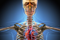
Last year the U.S. Armed Forces Institute of Pathology (AFIP) began using whole-body multislice CT as part of its postmortem examination of soldiers arriving at Dover Air Force Base in Delaware. CT is the newest component of a comprehensive analysis that aims to reveal not only how each service member died, but to learn as much as possible from the deaths, and perhaps even prevent some casualties.
The data might lead to a better helmet design, for example, or a better understanding of which injuries are survivable.
"What happens when a service member dies? First we get legal authority to do an autopsy and under federal law ... the armed forces medical examiner's system has the opportunity to perform autopsy or forensic investigation for any service member who dies of unnatural causes or is killed," said Dr. Lisa Pearse, MAJ, MC, from the medical examiner's office of the Armed Forces Institute of Pathology in Rockville, MD.
The workload has climbed with the death toll in the conflicts in Iraq and Afghanistan. Investigators have a backlog of research work that needs doing, Pearse said. But the needs of grieving families are a priority; the 24-hour investigative time limit is honored.
As a result, the one-day layover at the Charles C. Carson Center for Mortuary Affairs, part of the Dover Air Force Base complex, is packed with tests. Fingerprinting, autopsy, forensic pathology, and toxicology are performed along with 16-slice CT.
The bodies keep coming -- some 900 a year, close to 2,000 since the CT scanning component began in 2005 -- and they tend to be in bad shape. The enemy's reliance on attacks with explosives, in addition to helicopter crashes and other violently traumatic events, leaves only about 40% of the corpses intact. The other 60% come in pieces. Most are contorted due to rigor mortis; some are so twisted they don't fit into the CT bore, Pearse said.
"Most of these folks are not just dead -- they are way dead," Pearse said at the Society for Medical Innovation and Technology (SMIT) meeting in May. "No legs, no arms, no head, charred. It's not the standard sort of autopsy that you'd have in downtown Baltimore. These folks sometimes have an awful lot of damage to them, so absent (anatomy) becomes a burden."
The forensic examination is "a new procedure and a new program," she said. "If you go back to the first Gulf War, they only autopsied about one in five. Go back to Vietnam and there are even fewer."
The comprehensive exam was made possible by the opening of the 70,000-sq-ft Charles C. Carson Center for Mortuary Affairs in October 2003. According to Pearse, Congress members trudging through the old facility after the 2001 terror attacks weren't pleased with the poor condition and lack of capacity at the mortuary. In a rush to improve facilities after the events of September 11, the state-of-the-art replacement mortuary was completed in 18 months at a cost of $30 million, and is twice the size of the previous facility.
Since 1955, the remains of more than 50,000 service members have passed through the Dover base for identification and funeral preparations. At the old facility, the focus was on preparing the remains of fallen U.S. servicemen and women, as well as government officials and their families stationed abroad in Europe and Asia.
When a body arrives at the new Carson Center the investigation is more rigorous, starting with a baggage scanner that looks for unwelcome by-products.
"Literally, it's the airport baggage scanner," Pearse said. "What we're looking for is unexploded ordnance, bullets, hand grenades -- anything that might blow up on us. We do bar coding of the bodies and of the pieces that come with the bodies."
Intake photography documents the casualties, followed by fingerprinting, forensic pathology, digital x-ray, and eventually CT. Pathologists collect and examine bullet fragments.
The scan
The CT scan takes about 15 minutes, and at the outset there were concerns that full-body scans would slow down the investigation process unacceptably, Pearse said.
The bodies are scanned at 16 x 5-mm collimation, pitch of 1.75:1, 0.50-sec rotation speed, and table speed of 35 mm per rotation on a 16-slice CT scanner (LightSpeed 16, GE Healthcare, Chalfont St. Giles, U.K.). Images are retrospectively reconstructed at the CT console to a section thickness of 1.25 mm before being sent for review on a GE Advantage workstation, according to a recently published study on the group's research.
Standard protocols, such as a fragment protocol, have been developed to sidestep the development of a protocol for each individual case.
"We've developed protocols for fragment analysis, and we're working on a variety of other protocols," Pearse said in her talk. "Trying to get the data out has been one of our challenges, and it's going to be one of the big focuses for the next stage of this process."
This means implementing PACS, RIS, and data transmission capabilities to handle the high-volume workload, and being able to quickly send critical data to the end users of the information.
For image analysis, a GE software application called "Stickman" helps fill in data gaps from missing body parts by aligning key joints in the body, she said. Parts are color-coded: green is for normal intact anatomy, yellow is injured, and red signifies an absent body part.
The software is a step toward registration of the anatomy, will be required for the eventual move to robotics for removing bullet fragments and other tasks. But when too much anatomy is missing, "sometimes you reach the limits," where the software is unable to align and reconstruct the anatomy, Pearse said.
The user interface also counts fractures, tissue injuries, and metal fragments by size. This information is part of the scan and can be directly linked to the database, Pearse said, noting that it is meant to be a catalog rather than a comprehensive database.
A recent research project used CT head scan information to determine whether the correctly sized helmets have been issued to military personnel. "End users of the data include body armor developers, vehicle designers, military intelligence workers, and the trauma surgeons," Pearse said.
One interesting process to come out of the research has been metal analysis.
"Early on in the program our big concern was metal artifact, and a general effort was focused on getting rid of the metal artifact so that we could see the other things," she said.
But in the process of eliminating the artifacts, the researchers found they could identify different metal densities, "copper versus lead versus steel, which is something we never dreamed we would be able to do," Pearse said. "We're still struggling because it's so far out of the range of densities the CT scanner is designed to look at that we're having a little trouble differentiating it all, but the potential is there and I think it can be tweaked."
And as other investigators have found, bodies can quickly fall apart during a real autopsy. A CT scan acquired in advance permanently preserves the anatomy exactly as it was presented to the researchers, Pearse said.
Studying bullet tracks
Another project sought to advance the understanding of CT tracking of bullet wounds. Pearse and hear colleagues recently published a retrospective study on the role of multidetector-row CT (MDCT) in the forensic evaluation of gunshot wound victims (Radiology, August 2006, Vol. 240:2, pp. 522-528).
The study included 13 consecutive male wound victims (mean age 27) who underwent scanning with 16-slice MDCT prior to routine autopsy. The whole-body scans were interpreted on a 3D workstation (GE Advantage) by radiologists blinded to the autopsy findings. The researchers evaluated the findings for lethal wounds, number and location of wound tracks, injured structures, and the location of metal fragments. After image interpretation, the findings were validated by comparison with autopsy reports and photographs.
"Multidetector CT aided in correct identification of all lethal wounds, and metallic fragment location was always precise," the authors wrote. "In four cases, multidetector CT aided in accurate assessment of organ injuries and lethal wounds but led to underestimation of the number of wounds if co-mingling paths occurred. In two cases of a chest wound, multidetector CT aided in accurate assessment of the chest as having the lethal wound but failed to help identify specific sites of hemorrhage. In two cases of craniofacial injury, the path of the wound was not clear."
 |
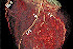
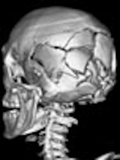
| Features of a gunshot wound to the head at virtual autopsy. Top and below, transverse CT images of a lethal gunshot wound to the head show multiple calvarial fractures, posterior settling of the brain, and pneumocephalus. Metallic fragments and foci of gas are present in the left cerebellum (below). A distinct linear bullet track is not present. Bottom, 3D volume-rendered image of the skull shows the comminuted fracture of the posterior skull. Republished with permission of the Radiological Society of North America from Radiology (August 2006, Vol. 240:2, pp. 522-528); published online before print as 10.1148/radiol.2402050972. |
 |
 |
Of 78 wound tracks, 10 (13%) were not seen at MDCT, the group reported. The missed tracks included six upper extremity wounds and four thigh wounds. In two cases, however, findings missed at autopsy (fracture of the cervical spine, bullet fragments in the posterior area of the neck) were identified at MDCT.
"We have shown that virtual autopsy with multidetector CT can aid prediction of lethal wound and fragment localization in victims with high-velocity gunshot wounds," the authors concluded. "Single gunshot wound tracks are well-defined. The major limitation occurs in classifying and identifying intersecting and co-mingling tracks."
High-volume challenges
"I'd love to say we haven't had any problems along the way, but it's not quite been the case," Pearse said in her SMIT talk. "The first thing that hit us was that the table length for a CT scanner is 4 feet 9 inches -- and we have very few soldiers that are 4 feet 9 inches."
The shortfall means that most cases have required two scans, doubling the time needed to complete them, she said. At the time of her talk, GE was in the process of providing a new table that is 6 feet 2 inches.
Then there is scanner capacity, or rather a lack thereof.
"The tubes for CT scanners are really not designed for (imaging) whole bodies over and over and over again," Pearse said. "What we find is that after we've done five or six bodies in a row we start getting degradation. The images are not as clear and actually they're very, very difficult to interpret."
This problem has been addressed by limiting the number of scans to four or five at a time. Subjects are often sent for odontology, fingerprinting, or some other process until the tube cools down.
"We've burned out tubes, and the tubes are hideously expensive," she said. "Thankfully there's a warranty, but it's just not what the system is designed to do. It's designed to image a head or designed look at a heart or a thorax. It's put a strain on the system."
The image files are huge, of course, and the network issues are just now starting to be addressed so that everyone who needs the files can access them, Pearse said. To that end, a massive effort is now under way to pull all the data sources together and make them accessible to all the end users. This raises a couple of other issues, such as medical privacy and military intelligence concerns, but these are also being addressed, she said.
"We've done sneakerware on disks from place to place. We're still working on data management and storage systems," she said.
CT's limitations
As for the modality itself, CT has its strengths and weaknesses in forensic pathology, Pearse said. It's excellent at seeing pelvic damage in particular, as well as vertebral fractures that are often missed at pathology, she said. But its limitations in soft-tissue imaging are a drawback, and many soft-tissue injuries are missed.
"We're trying to look for blast trauma and I don't think we're seeing a lot of blast injuries," Pearse said. But the biggest single mistake may have been a lack of planning for radiologists on the project.
"This plan was conceived by surgeons and executed by forensic pathologists," she said. "Nowhere in our original plan did we think of putting in radiologists -- and that was just a profound error on our part. We've gone back and rectified that and found some radiologists. Forensic pathologists don't have imaging expertise, and I suspect most surgeons don't either...."
It turned out the radiologists had their own catching up to do.
"Radiologists are used to ruling out 'X,'" Pearse said. "They were lost when we presented them with a whole-body CT scan and said, 'Tell us the problem.' It's taken them several months to get that overview (orientation) to tell us what's wrong." At first they were missing major fractures, for example, by simply failing to look for them, she said. But with time, practice, and better databases, their abilities have improved markedly.
To do the job correctly, the radiologists really needed a database with an interpretive interface, "so we could pull out all the scans with, say, metal in the head," Pearse said.
Current efforts are aimed at classifying and compiling the data in more sophisticated ways, and building the infrastructure to get it to the end users quickly -- all while avoiding conflicts with medical privacy and military intelligence issues, which are being addressed concurrently, Pearse said.
The next phase will incorporate robotics into the practice -- first for use in tagging and eventually for fragment retrieval, she said.
The AFIP's virtual autopsy program has been extraordinarily successful, commented Dr. Richard Satava, a professor of surgery at the University of Washington in Seattle who oversees the five Defense Advanced Research Projects Agency (DARPA) projects, including the virtual autopsy program, for the U.S. military.
"This is probably one of the most sobering projects that we have.... It brings to light not only medical issues but moral ethical and philosophical issues," he said following Pearse's talk.
Satava also saw great potential in the use of crossover technologies between the DARPA programs, such as the capture and classification of data on battlefield injuries. "When an injury occurs, we can interpret what happened (and learn) whether the injury is survivable or not," he said.
By Eric Barnes
AuntMinnie.com staff writer
August 3, 2006
Part I: Radiologists prepare for growing CT role in virtual autopsy, May 22, 2006
Related Reading
Diagnosis murder: Imaging as a psycholegal defense tool, June 29, 2006
Trauma Pod brings robotic ICU to the battlefield, June 26, 2006
Expanded role envisioned for imaging in autopsies, May 15, 2006
Image guidance evolves into imaging therapy, May 12, 2005
Copyright © 2006 AuntMinnie.com