Pediatric orthopedic imaging patients who need surgery often receive more x-rays and CT scans than nonsurgical patients, and orthopedic surgeons should seek options to reduce radiation exposure, according to research being presented this week at the American Academy of Orthopaedic Surgeons (AAOS) 2017 annual meeting.
Researchers from NYU Langone's Hospital for Joint Diseases performed a literature review of the different options in imaging technologies for pediatric orthopedic injuries and then quantified the amount of radiation in these scans. They found that patients who require surgery for hip dysplasia, scoliosis, and leg-length discrepancy are more likely to undergo imaging such as x-ray or CT.
As a result, these patients face a slightly higher lifetime risk for cancer and genetic defects, according to senior research author Dr. David Godfried and fifth-year resident Dr. Ayesha Rahman. They shared their findings in a scientific exhibit at AAOS 2017 in San Diego.
For example, pediatric patients with hip dysplasia that required surgery received two times more x-rays and underwent multiple CT scans, compared with pediatric patients who didn't undergo surgery. This radiation exposure cumulatively increased their overall risk of fatal cancer or genetic defects by less than 1% -- a small but significant risk, according to the researchers.
What's more, female scoliosis patients received two times more x-rays than nonsurgical patients, amounting to twice the radiation exposure to the breasts, ovaries, and bone marrow. This level of radiation exposure correlated to a more than 2% increased lifetime risk of fatal breast cancer, as well as an almost 1% higher risk of fatal leukemia and 3% greater risk of genetic defects. In comparison, nonsurgical patients had approximately half that risk, according to the team.
After reviewing the evidence, the researchers developed a list of best practices for orthopedic surgeons:
- Follow the ALARA, or "as low as reasonably achievable," principle to limit exposure to parts of the body that are absolutely essential for diagnosis.
- Eliminate repeated exposures resulting from technical errors.
- Limit precise collimation to the region of interest.
- Limit fluoroscopy to short bursts as needed; don't "go live."
- Utilize low-dose CT protocols that are adjusted for the size of the patient.
- Limit CT scans of the spine and pelvis in pediatric patients.
- Female patients are more susceptible to adverse effects of radiation than male patients.
- Scoliosis patients should have limited follow-up x-rays.
- Leg length, scoliosis, and hip dysplasia (anteversion) studies should utilize EOS Imaging's orthopedic digital radiography (DR) systems, rather than traditional x-rays.
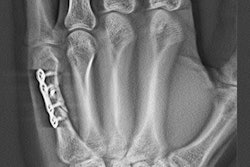
- X-rays are an acceptable diagnostic tool for extremities, such as the wrist, ankle, etc.
- CT scans are an acceptable diagnostic tool for triplane fractures.
The researchers said their institution has implemented several of these practices in collaboration with institution-wide efforts by the department of radiology to reduce radiation exposure.
"We have examined our use of x-rays in different clinical situations and the effect on patient outcomes, and have been able to reduce or eliminate the need for x-rays in many instances including certain postoperative and routine follow-up visits," Rahman said in a statement. "While x-rays are still a necessary and important diagnostic tool in the pediatric population, our goal is to reduce radiation exposure to these patients wherever possible without compromising patient care."
For example, children with scoliosis or suspected spine problems are now often imaged at NYU Langone with EOS orthopedic DR systems, which the group said provides useful information with about one-tenth the radiation exposure of a conventional CT or x-ray of the spine.
As for hip dysplasia cases, orthopedic surgeons at NYU Langone work with musculoskeletal radiologists to reduce a hip CT scan from an average of five to 10 slices to only one or two, according to the researchers. In addition, orthopedic surgery and radiology collaborators have implemented intraoperative use of low-dose protocols on fluoroscopy systems, which reduces radiation exposure to patients as well as physicians and staff in the operating room.