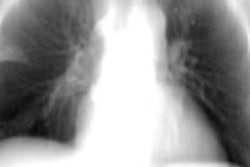
Characteristics seen on CT chest scans can enable physicians to distinguish between lung nodules caused by fungal infections such as valley fever and those caused by lung cancer, according to a poster presented at the recent American College of Chest Physicians (ACCP) annual meeting in Montreal.
With growing numbers of patients being screened for lung cancer using CT, more nodules are being detected on scans. But not all lung pathology is cancer, noted Dr. Reza Ronaghi, an internal medicine physician at the University of California, San Francisco, who presented the data.
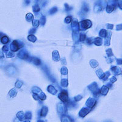
In fact, some nodules could be due to conditions such as coccidioidomycosis, or valley fever, a fungal infection that occurs in the southwestern U.S. The similarity of coccidioidomycosis to lung cancer can be challenging for physicians, so Ronaghi's team developed a calculator based on CT findings to help physicians tell the difference.
Stratifying nodules
Existing calculators employ clinical and radiographic criteria to risk-stratify nodules linked to lung cancer, but these calculators offer insufficient specificity to discriminate between nodules caused by lung cancer and those caused by coccidioidomycosis, Ronaghi explained.
Travelers to the southwestern U.S., or other geographic areas where the fungus coccidioides is commonly found, such as northern Mexico, can develop this lung infection, so clinicians based in areas not endemic to the fungus may encounter these patients, he noted.
Performing a biopsy on patients with this fungal infection is not required and, moreover, subjects them to unnecessary risk, Ronaghi stressed.
"These are patients who just need to be given antifungal therapy and then need to be followed," he said. "There are risks with undergoing biopsy, such as the complication of pneumothorax and bleeding after biopsy."
Indeed, Ronaghi and colleagues presented data at the American Thoracic Society (ATS) annual meeting in 2014 on complication rates in a population of patients who underwent CT-guided biopsies. They found that up to 24% of patients can develop pneumothorax, and 1% of patients can develop bleeding subsequent to a lung biopsy.
"We wanted to know if there is a way we can differentiate these nodules and avoid putting patients at risk [of complications of a biopsy]," Ronaghi said. "Is there a way we can separate out these patients [with coccidioidomycosis] and avoid them going through an unnecessary procedure?"
Ronaghi and colleagues retrospectively reviewed the records of 1,100 patients who presented with lung nodules between December 2009 and June 2013. A total of 302 patients had a confirmed diagnosis of either lung cancer (192 patients) or coccidioidomycosis (110 patients).
The investigators deidentified the CT scans of the patients and asked two chest radiologists, blinded to the results, to read the films, employing a standardized system for data collection. The findings were subsequently correlated with the patients and analyzed.
When the researchers compared features between the patients with lung cancer and those with coccidioidomycosis, some emerged as key to differentiating between the two conditions. There was no difference in terms of nodule density or calcification, but the size of the nodules differed: Nodule diameter was 4.2 cm in the lung cancer patients, compared with 2.9 cm in coccidioidomycosis patients (p = 0.0001).
The presence of chronic lung disease on CT was more often correlated with lung cancer nodules than coccidioidomycosis nodules, at 66% versus 19%, (p = 0.0001). Satellite lesions were more common in coccidioidomycosis patients, at 59% versus 15% (p = 0.0001). Nodule borders were also dissimilar between the two groups: 45% of patients with lung cancer had spiculated borders, compared with only 22% of coccidioidomycosis patients (p = 0.001).
"When the border [of the nodules] was smoother, [the diagnosis] was more likely to be coccidioidomycosis," Ronaghi said.
In terms of nodule pattern, 25% of nodules linked to the fungal infection were cavitary, while only 8% of lung cancer nodules were cavitary (p = 0.0001). And while it was not as statistically significant as other features, mediastinal adenopathy was more common among cancer patients, at 62% versus 57% (p = 0.035).
"When the patient's lymph nodes were enlarged, the diagnosis was more likely to be cancer," he said.
Ronaghi and colleagues incorporated the findings into a calculator designed to predict which patients with lung nodules have coccidioidomycosis and which have lung cancer. In addition to clinical features, the tool includes the patient's age, occupation, and whether he or she is a smoker as predictors.
"We have been studying this tool in our practice, and it is doing a good job," Ronaghi said, adding that decreasing the number of biopsies is a cost-saving measure to the healthcare system.
The researchers plan to present more data on the utility of the calculator at the ATS 2016 annual meeting, as well as share preliminary data from a multicenter trial examining the specificity of the prospective use of the calculator at next year's ACCP meeting.