Iterative reconstruction allows steep CT radiation dose reductions in urolithiasis imaging without loss of image quality or diagnostic confidence, researchers from Massachusetts General Hospital have found. The results may help allay fears of potentially excessive radiation in these oft-repeated scans.
In 25 patients undergoing ultralow-dose CT for suspected urolithiasis, the study team boosted image quality significantly by adding iterative reconstruction -- all while cutting the dose by about 85% with the ultralow-dose protocol, the group reported in a study published online in Radiology (August 13, 2012).
"Use of our modified scanning protocol and application of the [adaptive statistical iterative reconstruction (ASIR)] technique can enable substantial radiation dose reduction without introducing image-quality concerns for urinary stone diagnosis," wrote Dr. Naveen Kulkarni, Dr. Dushyant Sahani, and colleagues.
Urolithiasis is common condition, with a lifetime incidence of 5% to 10% in the U.S. that may be increasing over time. Recurrence in as many as 75% of those individuals is also a problem, according to the authors.
The prospect of some patients needing several scans over a lifetime to check for new stones has raised concerns that full-dose CT could be harmful in some cases, driving the search for lower-dose alternatives.
Those alternatives aren't likely to stray to another modality, however. These days, the traditional alternatives of radiography and ultrasound have been largely replaced by CT because of the latter's high sensitivity, which approaches 100% for stone detection, among other reasons, Kulkarni and his team wrote.
Typical low-dose CT protocols often increase image noise unacceptably, so a different kind of solution is called for, according to the authors.
The team sought to evaluate the performance of CT at 80 kV and 100 kV with 75 to 150 mA and the effect of ASIR (GE Healthcare) versus filtered back projection (FBP) on CT image quality in patients with urinary stone disease. All 25 patients (mean age, 33.5 years) received modified-protocol scans on a 64-detector-row CT scanner (Discovery CT750 HD, GE).
 |
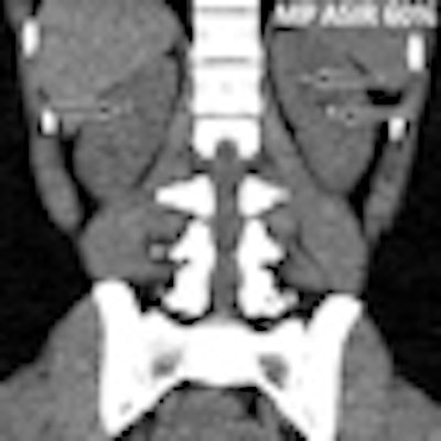
| Coronal CT images acquired in a 28-year-old man (body weight, 73 kg) with renal stone disease. Left (A) baseline reference-protocol (RP) CT image shows two 2- to 3-mm calculi in the right kidney. Right (B) follow-up modified-protocol (MP) CT image obtained six months later and reconstructed with 60% ASIR demonstrated two new tiny stones in the left kidney and a single calculus in the right kidney. The patient probably passed the inferiorly located calculus, which was not visualized at follow-up CT. The tiny renal calculi are depicted equally well on ASIR-reconstructed images and baseline CT images. Images republished with permission of RNSA from Radiology. |
In 13 of the 25 patients, prior standard-dose scans had also been performed with a 16-detector-row CT scanner (LightSpeed 16, GE) and a conventional FBP reconstruction technique. These 13 patients, who served as a reference protocol group for the study, had been scanned with a tube voltage of 120 kV and patient-weight-based tube-current modulation with a tube current range of 75 mA to 250 mA. The noise index was set to 25.
In the modified protocol, the tube voltage was cut from 120 kV to 80 kV for patients weighing less than 200 lb (and to 100 kV for those weighing more). The automated tube-current modulation technique was set to 75 mA to 150 mA with a noise index of 30 for both groups. The researchers created one FBP and two ASIR (60% and 80%) image sets.
Two readers independently examined the FBP and ASIR datasets for subjective image quality on a scale of one to five, noise on a scale of one to three (3 = excessive noise), and diagnostic confidence on a scale of one to three (3 = highly confident). They also documented the number, size, and location of stones.
The results showed overall low-quality images for the modified protocol at FBP reconstructions (mean image quality, 2.5), but there was significant improvement with ASIR reconstruction (mean image quality, 3.4; p = 0.03).
Even using the modified protocol, however, all 33 stones in 17 patients (mean diameter, 6.1 mm; range, 2-28 mm) were diagnosed by both readers, whether reconstructed with FBP or ASIR.
In 20 (80%) of 25 patients, ASIR-reconstructed images were also rated adequate for making additional diagnoses in the abdomen (image quality, 2.0). FBP-reconstructed images had an image-quality score of 1.3, however.
The overall radiation dose for modified-protocol CT was 1.8 mGy, which combined patients weighing less than 200 lb (1.3 mGy) and patients weighing more than 200 lb (2.3 mGy). In comparison, the dose was 9.9 mGy (p = 0.001) in the 13 reference-group patients.
For detecting stones using the modified protocol, the diagnostic confidence was higher for 60% ASIR images (mean score, 3.0) and 80% ASIR images (mean score, 3.0) than for FBP images (mean score, 1.9; p = 0.03).
Beats other dose-reduction techniques
"CT examination with a modified protocol and use of ASIR provides diagnostic-quality images that can be used for evaluating urinary calculi at a substantially reduced radiation dose," Kulkarni and colleagues wrote.
Previous studies have shown that performing reduced-kV urolithiasis scans using FBP imaging leads to substantial degradations in image quality, they wrote. In this study, 80% of the images acquired with the modified protocol and FBP reconstruction had suboptimal image quality.
"In addition to urolithiasis detection, it is also imperative to maintain acceptable image quality for the identification of alternate diagnoses in patients who do not have urinary tract calculi," the group added.
The study's limitations included the lack of a reference-standard image for each calculus, the use of a vendor-specific iterative reconstruction technique that is not the standard of care, and the lack of patient randomization to CT reconstruction techniques.
However, image quality can be expected to continue improving over time as more facilities begin to use iterative reconstruction packages from major CT manufacturers that have recently been approved by the U.S. Food and Drug Administration (FDA), the group noted.
It is important to emphasize that for patients with lower body weight, the actual absorbed organ radiation dose could be higher even when the same scanning parameters are used, whereas in heavier patients, the absorbed organ radiation dose can be lower, according to the researchers.
"CT radiation dose reduction to 1.8 mGy can be achieved with ASIR image reconstruction for evaluation of urolithiasis, while maintaining acceptable image quality and diagnostic confidence comparable to that with CT protocols with a radiation dose of 9.9 mGy," Kulkarni and colleagues wrote.