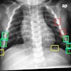
Applying certain types of postprocessing techniques to digital radiography (DR) chest images can help increase lung nodule detection when the images are analyzed by computer-aided detection (CAD) software. But the performance improvement comes at a price of more false positives.
In a new study from China, researchers at Capital Medical University's Beijing Friendship Hospital applied different types of image processing techniques to DR images, then analyzed the impact of the techniques on CAD performance (Journal of Digital Imaging, February 1, 2008).
The growth of DR is creating new possibilities for digital image enhancement and analysis, with users able to manipulate various structures and spatial frequencies in digital data. At the same time, DR images can be easily input into CAD software for analysis, an important consideration if the modality is to ever develop a role in lung screening of large patient populations. But few studies have examined the impact of image postprocessing on CAD results, according to the researchers.
The study examined 116 lung cases collected on a DR system (Digital Diagnost, Philips Healthcare, Andover, MA), divided evenly between normal cases and those with noncalcified nodules up to 15 mm in diameter. The group discarded cases with very small nodules that weren't detectable on DR, those with nodules larger than 15 mm, or cases with more than five nodules. DR images and CAD results were reviewed on 2K x 2K display monitors (E-2320, Barco, Kortrijk, Belgium), and cases were confirmed via consensus reading of two radiologists reviewing chest CT studies.
Both normal and abnormal cases were processed using the Unique image processing algorithms available with Philips DR systems. Three types of image processing protocols were used: standard images (no special processing), a high-pass algorithm, and a low-pass algorithm. After processing, images were analyzed with a commercial CAD application (IQQA-Chest, version 1.2, EDDA Technology, Princeton Junction, NJ).
The high-pass and low-pass algorithms, also known as multiscale processing, offer different methods for analyzing structures in digital images. The high-pass algorithm enhances higher spatial frequencies, such as the edges of lung nodules and noise. As a result, the contrast between a pulmonary nodule and its surroundings is lower, the researchers stated.
On the other hand, a low-pass filter enhances lower spatial frequencies, enhancing the contrast between a pulmonary nodule and its surroundings. In addition, normal anatomic structures in the lungs, such as pulmonary vessels, are enhanced.
Each of the different image processing algorithms resulted in different levels of performance by the CAD software. The chart below demonstrates the differences:
|
||||||||||||||||||||
"Among the three types of images reviewed, the low-pass images ... appear to improve the accuracy of lung nodule detection with the CAD system, whereas the false-positive rate is highest among the three types of the postprocessed images," the researchers wrote.
One possibility is that the low-pass algorithm enhances both pulmonary nodules as well as normal anatomic structures in the lungs, such as pulmonary vessels, producing a higher detection rate but also more false positives. Meanwhile, the high-pass algorithm produced lower contrast between a pulmonary nodule and its surroundings, which could account for the lower detection rate.
The results showed that different image processing techniques can affect CAD performance, but the study did not address the question of whether the effect "carried over to the combined performance of physicians together with CAD," the researchers wrote. They recommended further research to address this question.
By Brian Casey
AuntMinnie.com staff writer
February 12, 2008
Related Reading
Chest x-ray lung CAD reimbursement picks up, October 24, 2006
X-ray CAD aids in early lung cancer detection, June 22, 2006
NCI CAD database program gathers momentum, March 16, 2006
Chest x-ray CAD reaches reimbursement milestone, November 22, 2005
X-ray CAD finds bone defects in rheumatoid arthritis, May 18, 2004
Copyright © 2008 AuntMinnie.com
