A new 4D PET reconstruction method can improve cardiac PET image resolution by compensating for both respiratory and cardiac motion, according to researchers from Tsinghua University in Beijing and Johns Hopkins University.
Fully compatible with current imaging protocols, the method also reduces the noise level of cardiac-gated PET images by using all of the PET data to reconstruct every cardiac-gated image, according to presenter Si Chen of Tsinghua University's department of engineering physics.
"Our method can significantly improve the image quality of cardiac PET imaging, i.e., enhance image resolution and reduce image noise level," Chen told AuntMinnie.com. "This will likely benefit the accuracy of physicians' interpretation of the PET images."
Chen presented the 4D PET reconstruction method during a presentation at the recent Society of Nuclear Medicine and Molecular Imaging (SNMMI) annual meeting in Miami Beach, FL.
Patient respiratory and cardiac motion can blur cardiac PET images generated by current standard PET imaging protocols and reconstruction methods. While attempts have been made to freeze either respiratory or cardiac motion in cardiac PET imaging, most of the proposed methods compromise the image noise level by throwing away some data to deal with the blurring effects of patient motion, Chen said.
To improve this situation, the research team developed a 4D PET image reconstruction method that first analyzes the PET list-mode data to extract the respiratory motion signal. This signal is used later for respiratory gating.
Next, the method estimates patient respiratory motion based on the respiratory-gated PET data. The previously extracted respiratory signals are combined with the electrocardiogram (ECG) triggers that were recorded during the PET scan to achieve cardiac and respiratory gating for the list-mode data, Chen said.
Finally, the 4D reconstruction method is applied to the respiratory and cardiac-gated PET data, yielding reconstruction of cardiac-gated PET images that include compensation for respiratory and cardiac motion.
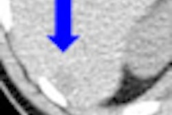
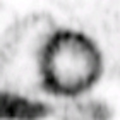
To test the technique, the group applied it to two PET stress studies. One was a nitrogen-13 ammonia stress myocardial perfusion of a 57-year-old female with a healthy heart, while the other was an F-18 FDG myocardial viability study on a 34-year-old male with a diseased heart. After the reconstruction method was performed, the images were evaluated by physicians from multiple centers, Chen said.
 |
| All images courtesy of Si Chen. |
 |
The researchers found that the reconstruction method yielded a 15% improvement in myocardium-to-chamber contrast compared to the conventional method that did not use any motion compensation. Furthermore, the noise level of each 4D PET image was 60% lower than with cardiac-gated images reconstructed using the conventional method; therefore, the cardiac motion defect became more prominent with the new method, according to the researchers.
"Improved image quality implied improved diagnostic accuracy," Chen said. "This can be traded for the reduced scan time or less patient radiation dose [less injection of the radiotracer] with the same diagnostic accuracy."
Chen said the 4D reconstruction technology will be further validated and evaluated with more patient data over the next two years.