When pulmonary embolism (PE) is suspected in an emergency room patient, there's no time to waste. Offering equivalent diagnostic performance to a PACS workstation, Apple's iPad just might be the tool to speed diagnosis, according to researchers from Johns Hopkins School of Medicine.
In a study published online in Emergency Radiology, a Johns Hopkins team found no difference between the use of an iPad and a PACS workstation in diagnostic accuracy for detecting PE. As a result, the popular tablet offers potential for applications such as after-hours reading or for an attending consult from a remote location, lead author Dr. Pam Johnson told AuntMinnie.com.
Seeking to evaluate the performance of the iPad as a mobile image interpretation tool, the research team prepared a study that included 50 deidentified contrast-enhanced CT exams that were performed for suspected PE and had been compiled as a learning tool to prepare residents for night-call coverage (Emergency Radiology, March 26, 2012). In the set of 50 exams, there were 25 PE cases, including easier diagnoses, such as large main pulmonary emboli, and harder ones, such as subsegmental emboli.
Both thick-section (3-mm section thickness x 3-mm interval) and thin-section (0.75-mm section thickness x 0.5-mm interval) reconstructions were sent to the PACS after image acquisition. At Johns Hopkins, the protocol is to review the thin-section volume as axial sections on all patients who have no emboli apparent on the 3-mm sections, according to the researchers.
The thin-section volume can also be used to generate interactive multiplanar reconstructions (MPRs), according to the authors. Readers in the study interpreted the exams as axial sections, and utilized MPRs as necessary. On the iPad app (Syngo Web Viewer, Siemens Healthcare) readers can shift instantaneously between viewing axial sections, 2D MPRs, maximum intensity projections (MIPs), or 3D volume rendering, according to the researchers.
Two fellowship-trained junior radiology attendings read the CT exams; one read the studies first on the iPad and then, after at least two weeks, interpreted the cases again in a different order on a PACS workstation. The second attending followed the same protocol, but in the opposite order.
Both readers produced identical interpretation accuracy on both platforms.
Diagnostic performance, iPad versus PACS workstation
|
The differences were not statistically significant. The one PE that was missed on the iPad was due to reader error, as it was apparent on the iPad and identified by the second reader, according to the researchers.
 |
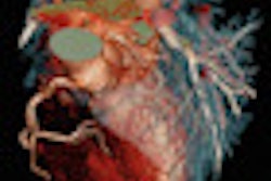
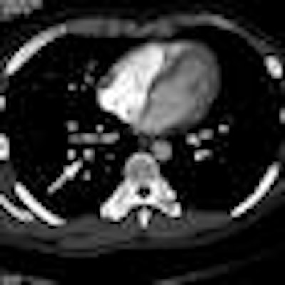
| Axial image (top) and sagittal multiplanar reconstruction (bottom) from contrast-enhanced MDCT viewed on the iPad. Arrows show right lower lobe segmental pulmonary embolism. All images courtesy of Dr. Pam Johnson. |
 |
Although interpretation speed wasn't measured during the study, the reviewers were surprised at how fast the iPad was, Johnson said. Image load time ranged from 700 to 1,000 slices per second at 512 x 512 resolution.
"Both readers commented that the iPad seemed like a faster method of display, and both selected the narrow reconstructed sections as primary means of review, generally in the range of 600-800 slices," the authors wrote.
The researchers acknowledged a number of limitations in their study, including the use of only two readers and a case mix that had an incidence of PE that's many times higher than in an inpatient or outpatient population. Nonetheless, the authors believe that the number of positive cases yielded better statistical data and allowed the residents to gain more exposure to the appearance of positive PE exams.
"The results of our study provide evidence of the utility of the iPad for CT display in patients with suspected pulmonary embolism and should serve as the basis for additional research investigations that focus on other emergency radiology CT applications," the authors concluded.