The iPhone is a fine platform for interpreting knee MRI images, and for most injuries it's equivalent to a workstation, according to a new study by Canadian researchers that was presented earlier this month at the American Academy of Orthopaedic Surgeons (AAOS) meeting in San Francisco.
Compared to arthroscopy, expert interpretation of iPhone images showed high sensitivity and specificity for medial meniscus and cruciate ligament injuries, though accuracy was reduced for lateral meniscus tears and cartilage injuries, said Dr. John Theodoropoulos, an orthopedic surgeon from the University of Toronto.
Several imaging applications for mobile devices have been created since the introduction of the Apple iPhone in 2007, including apps for acute appendicitis, acute cervicodorsal spine trauma, acute stroke, and coronary artery stenosis based on CT images. However, few studies have explored the interpretation of MRI images on the diminutive platform, Theodoropoulos and colleagues wrote in their AAOS poster presentation.
"Given that radiologists are not always at the hospital, they're at home, in different centers ... we were looking for ways to look at imaging outside the hospital, and we thought that doing this via the iPhone would be ideal [because] it's something people carry around with them all the time," Theodoropoulos told AuntMinnie.com in an interview. "The technology already existed, and we thought it was a good idea for sports medicine."
The DICOM viewer software used in the study (version 1.14, OsiriX) converts eFilm workstation images to DICOM images so they can be viewed on an iPhone, he said. The images can then be enlarged or adjusted for brightness, contrast, and other image properties, Theodoropoulos said.
Their prospective study examined the diagnostic performance of an iPhone application for the diagnosis of intra-articular knee pathology at MRI followed by arthroscopy in 50 cases throughout 2009. This is the first MRI iPhone study he's aware of, Theodoropoulos said.
The group obtained MR images of the knee at 1.5-tesla (Sigma Excite, version 12.0, GE Healthcare) using a standard knee coil. They acquired sagittal fast-spin echo (FSE) proton density (PD), sagittal FSE T2-weighted fat sat, high-resolution coronal FSE PD, and axial FSE T2-weighted fat sat sequences, all performed with 4-mm slice thicknesses and field-of-view of 14 x 14 cm. Theodoropoulos did all of the orthopedic surgeries, and recruited the patients for the study.
To establish interobserver variability, two musculoskeletal radiologists examined the randomly ordered cases on an iPhone and a conventional PACS workstation -- with the first examining all 50 cases, and the second reading 25 randomly selected cases. Interpretation was timed to the closest minute, and the same cases were read on different platforms no sooner than two months apart.
Handheld images were reviewed using version OsiriX viewing software on an Apple iPhone 3G. Workstation images were examined using eFilm workstation software version 3.0 (Merge Healthcare) and read on a flat-panel monitor. A single orthopedic surgeon who had performed all of the arthroscopy procedures reported the cases using the same standardized reporting template used by the radiologists, Theodoropoulos and colleagues reported.
Reviewing the iPhone images, the musculoskeletal radiologists "were blinded to what the surgery showed, and what the initial MRI showed" on the workstation, "and they independently assessed the imaging results, and we compared those to the surgical findings, and to the eFilm MRI," he said.
 |
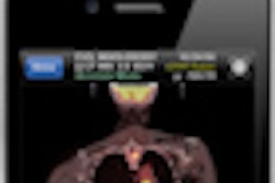
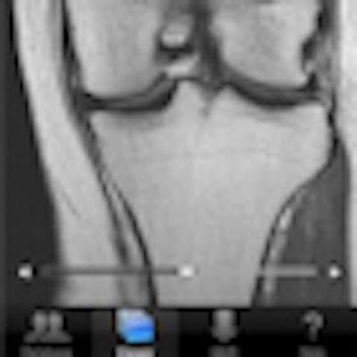
| All handheld mobile device images were reviewed using OsiriX DICOM viewer software version 1.1.4 on an Apple iPhone 3GS device. The iPhone 3GS display has a diagonal screen size of 8.9 cm and a screen resolution of 320 x 480 pixels. Image courtesy of Dr. John Theodoropoulos. |
The review focused on five major knee structures, including the medial and lateral menisci, anterior cruciate ligament (ACL), posterior cruciate ligament (PCL), and articular cartilage. Further subdivision of the menisci and cartilage produced 14 separate data points for review and comparison, which were graded on a scale of 0-2, with 0 being normal and 2 being a "definite tear" or other abnormality. Sensitivity and specificity were calculated versus arthroscopy as the gold standard.
iPhone interpretations showed high sensitivity and specificity for medial meniscus and cruciate ligaments injuries with lower sensitivity for lateral meniscus tears and lower specificity for cartilage injuries, the authors reported. And compared to much larger the PACS workstation interpretation on a flat screen, the iPhone showed excellent agreement for medial meniscus and cruciate ligament injuries and good agreement for cartilage injuries.
The improved accuracy for meniscus and cruciate ligament injuries versus cartilage problems on the iPhone interpretations mirrors what radiologists encounter on regular workstations, Theodoropoulos said.
"That's common," he said. "This mirrors what happens all the time clinically" because cartilage is not as well visualized on the workstation or the iPhone. Still, the iPhone app missed two cartilage tears versus the full-sized workstation, he said.
Accuracy of iPhone or PACS workstation for MRI
|
|||||||||||||||||||||||||||||||||||
Agreement between iPhone and PACS in interpretation of knee MRI
|
|||||||||||||||||||||
The use of a mobile device for the interpretation of knee MRI is promising, Theodoropoulos and colleagues concluded. Access to mobile interpretation "may increase access to subspecialty trained musculoskeletal radiologists making interpretation of musculoskeletal MRI available in remote areas without the need for fixed workstations," they wrote.
Theodoropoulos, who works with professional sports teams, said MRI scanners are common at professional sporting events; for example, one was in place at the recent Super Bowl.
"Maybe they'll have MRI machines at every NFL game going forward, but you probably don't need a radiologist on the sideline of every football game," he said. "You could just have a radiologist for the NFL sitting somewhere in an office where [reviewing the images on an iPhone] he could at least rule out major injuries and allow return to play."
And if an orthopedic surgeon reading the MRI images acquired at the stadium isn't sure about what he's seeing, he can always get a second opinion by forwarding the images to his radiologist friend with an iPhone, Theodoropoulos said.