Applying computer-aided detection (CAD) to digital x-ray images can help radiologists identify significantly more small lung nodules in screening radiographs for lung cancer, according to a study of asymptomatic adults in China.
Based on 328 digital radiography (DR) and 200 computed radiography (CR) screenings read with and without CAD, Yan Xu, PhD, and colleagues at Beijing Friendship Hospital - Capital Medical University in Beijing determined that CAD dramatically improved the radiologists' performance. Xu presented the results as a poster at the 2010 RSNA meeting.
The combined sensitivity of four radiologists for detecting small (5-15 mm diameter) pulmonary nodules rose from 65.6% when the digital images were read without CAD to 80.6% when they were re-read with CAD aiding the interpretation. A receiver operator characteristics (ROC) analysis also indicated a statistically significant improvement (p < 0.05).
CAD's sensitivity rates for studies performed with DR and CR were 81% and 79%, respectively. The use of CAD was accompanied by an average of two false positives per case.
 |
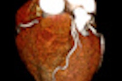
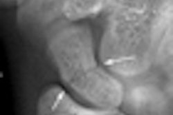
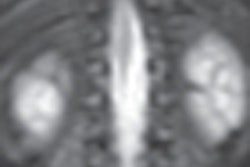
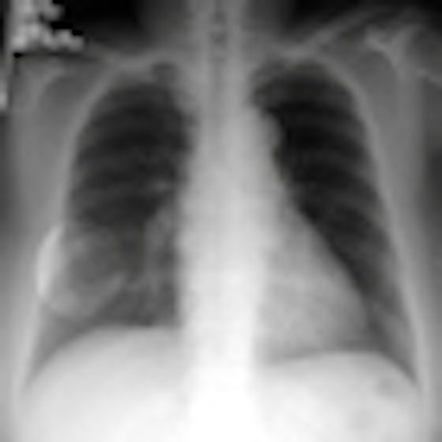
| CAD helps define the region of interest (ROI) harboring a subtle pulmonary nodule in a 79-year-old woman. (A) Arrow denotes nodule on x-ray. (B) CAD defines ROI. (C) CT indicates a small nodule in the middle lobe of the right lung. (D) A slide of sampled tissue from the nodule confirmed the presence of a well-differentiated adenocarcinoma. All images courtesy of Yan Xu, PhD. |
 |
Cases were drawn from an archive of 128,200 lung cancer screening exams of asymptomatic adults in China. The study group was equally divided between men and women (average age, 52 years; range, 20-86). Sixty-two percent were cigarette smokers.
The four radiologists reading for the study underwent CAD training involving six supervised cases, according to Xu. CR and DR image data were stored on the hospital's PACS. Images were viewed in a 2,000 x 2,000-pixel matrix and 12-bit grayscale format, and they were reviewed with commercially available CAD software (version 1.0, IQQA-Chest, EDDA Technology, Princeton, NJ).
Positive cases were initially identified by two chest radiologists, each having more than 15 years of diagnostic experience with pulmonary disease. They detected suspicious nodules from the selected sets of DR and CR studies and described their location, size, and probability for metastatic disease using CAD and corresponding CT scans for guidance.
During the study, the CAD algorithm generally performed poorly when assessing complicated lesions and benign abnormalities, such as pneumoconiosis and hematogenous pulmonary tuberculosis, Xu wrote in response to questions from AuntMinnie.com.
"The detected sensitivity of [CAD] will reduce in the practical application, so it should be improved," she wrote.
IQQA-Chest version 1.0 was awarded U.S. Food and Drug Administration (FDA) clearance for distribution in 2004. EDDA introduced version 2.0 of IQQA-Chest in November 2006.
By James Brice
AuntMinnie.com contributing writer
January 10, 2011
Related Reading
Chest x-ray CAD offers value in detecting lung cancers, June 10, 2010
Will new DR technologies offer alternative to CT? Part 2, March 25, 2010
CAD for chest x-ray detects overlooked subtle lung cancer, June 23, 2009
EDDA launches V2.0 of IQQA-Chest CAD software, November 20, 2006
Copyright © 2011 AuntMinnie.com