Studies have shown that MR-guided focused ultrasound (MRgFUS) therapy can be a practical, less invasive alternative to surgical resection for treating uterine fibroids. But MRI may have an additional role in this treatment protocol, helping clinicians assess at the beginning how useful the surgery will be in reducing fibroid volume and relieving symptoms.
Researchers at Brigham and Women's Hospital and Harvard Medical School in Boston and the Mayo Clinic in Rochester, MN, used MRI to distinguish predictors of success for MRgFUS surgery. Their findings appear in the October issue of Radiology (October 2008, Vol. 249:1, pp. 187-194).
Dr. Zsuzsanna Lénárd and colleagues conducted a retrospective analysis of 71 symptomatic fibroids in 66 women who had been treated with MRgFUS surgery between February 2002 and December 2005. These 66 women underwent complete follow-up, including fibroid volume and symptom assessment at six and 12 months post-treatment. The device used for MRgFUS surgery was a combination of a 1.5-tesla magnet (Signa, GE Healthcare, Chalfont St. Giles, U.K.) and a focused ultrasound device (ExAblate 2000, InSightec, Tirat Carmel, Israel).
Before treatment, the women were evaluated with MR imaging, including three plane localizer images; axial, sagittal, and coronal T2-weighted spin-echo images; T1-weighted spin-echo images; and multiphase fat-suppressed axial T1-weighted spoiled gradient-echo images. Patients also received contrast-enhanced multiplanar T1-weighted images. Lénárd's team used software to calculate the volume of treated fibroid and nonperfused volume (NPV).
At six and 12 months, each woman completed a questionnaire to determine her symptom severity score (SSS); the questionnaire used a five-point scale, with responses ranging from "not at all" to "a very great deal." Maximum symptom severity was represented by 100 points.
Before receiving MRgFUS surgery, the mean volume of the fibroids assessed was 255.5 cm ± 201.7, and the women had an SSS range of 61.5 ± 14.9.
At the six-month follow-up, the mean volume of the treated fibroids was reduced by 12.6% ± 16.9 compared with pretreatment data. Of the 71 treated fibroids, 55 (77.5%) had decreased in size, two (2.8%) had remained the same, and 14 (19.7%) had increased in size. The mean SSS was 34 ± 17.2.
At the 12-month follow-up, the mean volume of the treated fibroids was reduced by 9.3% ± 24.8. Fifty-three (74.6%) had decreased in size, one (1.4%) had stayed the same, and 17 (23.9%) had increased. All of the 18 women whose fibroids had not gotten smaller reported symptom improvement, however. The mean SSS was 37.6 ± 17.8.
Baseline characteristics of patients
|
||||||||||||||||||||||||||||||||||||||||||||
| *Numbers used to calculate percentages are in parentheses. †Values are mean ± standard deviation. Chart and all images courtesy of the Radiological Society of North America; from Lénárd Z, McDannold N, Fennessy F, et al. Uterine leiomyomas: MR imaging-guided focused ultrasound surgery -- imaging predictors of success. Radiology. 2008;249:187-194. |
||||||||||||||||||||||||||||||||||||||||||||
The researchers found that both pretreatment fibroid signal intensity (SI) and post-treatment NPV predicted that the fibroid's volume would have decreased by 12 months' follow-up: A decrease in fibroid volume correlated to the percentage of the fibroid that was nonperfused after MRgFUS.
"Fibroids with an NPV of at least 20% or with low SI both showed significantly larger volume reduction than fibroids with an NPV less than 20% or with high SI," Lénárd and colleagues wrote. "Patients whose fibroids demonstrated an NPV of at least 20% also experienced a larger decrease in symptom severity score than did patients with fibroids with an NPV less than 20%."
 |
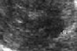
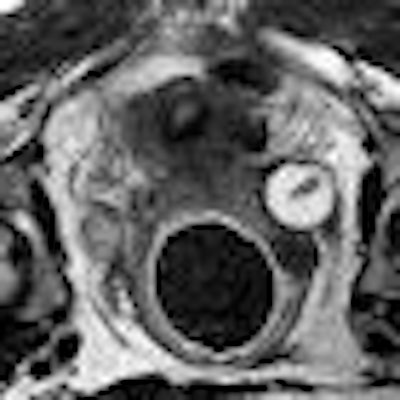
| Above, T2-weighted axial MR image of a hypointense fibroid in a 42-year-old patient before MR-guided focused ultrasound surgery. Below, T1-weighted gadolinium-enhanced axial MR image of the same fibroid immediately after treatment. The outlines of treated fibroid and NPV were contoured with electronic calipers to compute the area per section by using volumetric analysis software. |
 |
 |
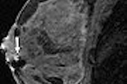
| Above, T2-weighted axial MR image of a hypointense fibroid in a 46-year-old patient before MR-guided focused ultrasound surgery. Below, T1-weighted gadolinium-enhanced axial MR image of the same fibroid immediately after treatment. |
 |
The study suggests that MRI can help predict a successful outcome of ultrasound-guided fibroid surgery, according to the researchers.
"Our findings may help both in selecting appropriate patients for MR-guided focused ultrasound surgery and in predicting patient outcome," they wrote.
By Kate Madden Yee
AuntMinnie.com staff writer
September 25, 2008
Related Reading
Study: MRI identifies submucosal fibroids that may migrate into endometrium, May 19, 2008
MR-guided ultrasound surgery of uterine fibroids is cost-effective, April 21, 2008
MRgFUS an option for many women seeking less invasive fibroid treatment, June 18, 2007
MRI-guided ultrasound surgery improves fibroid symptoms, June 1, 2007
MR signal, nonperfusion drive long-term success of MR-guided focused US therapy, May 20, 2007
Copyright © 2008 AuntMinnie.com