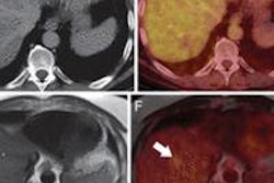
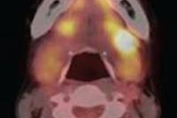
The use of PET/CT to create 3D images of a patient can help surgeons see detailed anatomical structures and gain better views of a tumor.
That finding comes from researchers at Thomas Jefferson University, who have developed a hologram-like display of a patient's organs that surgeons can use to plan surgery. The study was published online September 24 in PLOS One.
Matthew Wampole, PhD, from the department of biochemistry and molecular biology, and colleagues produced a surgical simulation of human pancreatic cancer reconstructed from a patient's PET and contrast-enhanced CT scans.
Six Jefferson surgeons evaluated the 3D model for accuracy, usefulness, and applicability to actual surgical experience.
The surgeons concluded that the 3D technique would help in planning an operation and would be most useful if it were accessible in the operating room during surgery. The 3D image is designed to speed the excision of malignant tissue and avoid bleeding from unusually located arteries or veins.