It's critical to diagnose injuries quickly and accurately in trauma cases. In sternal injuries, incorporating sagittal reconstructions in chest MDCT studies can help support that goal, researchers from the University of Alabama at Birmingham (UAB) concluded.
The UAB research team determined that evaluating the sternum in the sagittal plane increases injury detection as well as diagnostic confidence. Even better, these image reconstructions do not require additional radiation exposure for patients and can be automated to add little or no extra cost or time to the technical aspects of the imaging process, said Dr. Nina Terry, an assistant professor of radiology at UAB University Hospital.
"We argue that the time gained due to increased diagnostic confidence offsets any time spent on review of the sagittal images," she said. "Thus the sagittal images improve the diagnosis of sternal injuries over axial images alone with minimal additional monetary or time costs and no additional radiation."
Terry presented the UAB team's research at the American Roentgen Ray Society (ARRS) annual meeting.
Assessing patients with polytrauma requires rapid and accurate identification of injuries, and each additional test or imaging sequence takes up more time. However, it's justified if the tests significantly improve identification of serious injuries and confidence in the presence or absence of such an injury, according to the researchers.
Sternal injuries may be obscured on axial imaging due to the curved configuration of the anterior chest wall. That's a real problem because sternal injury is not only a direct source of pain, it can also indicate the presence of aortic or cardiac injury, according to the team.
Sagittal reconstructions
Believing that sagittal reconstructions could increase sensitivity and specificity for sternal and spinal injuries, in January 2009 UAB began to perform sagittal reconstructions on all of its body trauma CT cases (chest, abdomen, and pelvis). These were sent to the PACS along with the axial images and coronal reconstructions.
UAB's chest section then sought to quantitatively verify the initial perception that the sagittal images were improving sensitivity, specificity, and confidence in diagnosing sternal injuries and provide further support for their routine use, Terry told AuntMinnie.com.
The group's study was designed to assess intrareader agreement and reader diagnostic confidence with and without the use of sagittal reconstructions. The researchers retrospectively identified 52 trauma cases with sternal injury and an additional 113 patients without sternal injury to serve as a control group from a six-month period in 2009.
One sternal injury case was excluded from the study due to a lack of sagittal reconstructions, while 10 control cases were removed due to a lack of adequate sagittal images, motion on the examination, or a field-of-view that was too small, according to the researchers.
Five readers independently assessed deidentified sets of axial-only trauma chest CT images, and then, at a later date, a deidentified set of sagittal-only images of the same trauma chest CT. The radiologists recorded findings for the presence of a sternal injury (including secondary signs of sternal injuries such as adjacent hematomas), and assessed their confidence for the presence or absence of a sternal injury on a five-point scale.
The Cohen kappa statistic was used to assess the degree of agreement, and it was calculated for each of the 10 combinations of the five raters. Kappa coefficients ranged from 0.84 to 0.52, with a median of 0.60 for the axial MDCT images. The intraclass correlation for single-score reliability among a fixed set of raters was 0.64. Correlation between each rater's diagnosis of sternal injury and his or her confidence rating ranged from 0.64 to 0.97.
Better intrareader agreement
However, sagittal reconstructions generally yielded higher agreement indexes with kappa coefficients, ranging from 0.86 to 0.57, with a significantly higher median of 0.79 (p = 0.02) and a single-score fixed rater intraclass correlation of 0.75 (p = 0.03). The relationship between the raters' diagnoses and their confidence ratings were also generally higher with sagittal images, with correlation coefficients ranging from 0.82 to 0.89.
"The statistical results showed higher agreement between radiologists in the presence or absence of injury with the sagittal images," Terry said. "There was also higher confidence of the individual radiologist in their diagnosis when viewing the sagittal images."
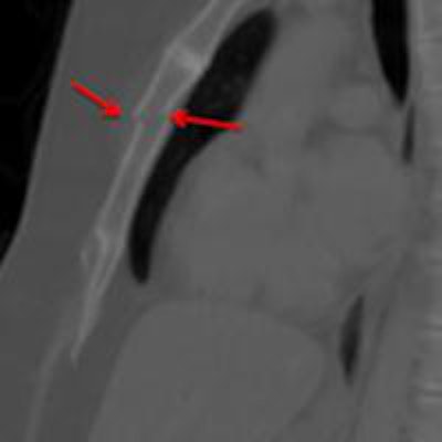
 Images courtesy of Dr. Nina Terry.
Although the sagittal images did lower disagreement over the diagnosis of sternal injury, they didn't eliminate it.
"We plan to review these cases to evaluate for potential causes of discrepancy such as breathing motion," she said.
In the next phase of the research, the UAB team plans to conduct additional statistical analysis of the data, including performing a receiver operator characteristics (ROC) analysis and a review of the correlation between the presence of presternal and/or mediastinal hematoma and the finding of fracture, Terry said.