Duke University Medical Center's Multi-Modality Imaging Lab (MMIL) is combining SPECT and CT technologies into a single device for 3D volumetric breast imaging to obtain both functional and anatomical information in one exam.
The SPECT/CT system, which uses a single gantry, is the brainchild of associate professor Martin Tornai and a group of students in the MMIL in Durham, NC. The novel hybrid imaging system was the topic of a presentation at the 2007 SNM annual meeting in Washington, DC.
The SPECT system itself is capable of full 3D motion and consists of a 16 x 20-cm2 area cadmium zinc telluride (CZT) detector with 2.5-mm pixelation and 6.7% full-width half-maximum energy resolution at 140 keV, as well as a high-resolution, parallel hole collimator.
The CZT detector, which detects emitted rays from radionuclides that are injected into the breast and torso, is coupled to a radius of rotation (ROR) stage and, in turn, is attached to a goniometer. The goniometer measures and monitors the angle of the camera, and allows for its rotation to a precise angular position.
As the beam passes through the uncompressed breast, it is detected by a flat-panel cesium iodide (CsI) detector. Unlike the SPECT system, the CT system, which uses a chromatic x-ray source, is restricted to a fixed tilt and can only move circularly around the breast.
|
Having trouble viewing this clip? Click here to view full-size clip or to change format. Video courtesy of Duke University Medical Center Multi-Modality Imaging Lab. |
Duke researchers use iterative reconstruction to obtain a 3D volume dataset of the breast. "By using external fiducial markers that are placed around the breast, we can fuse the two datasets together to show us a 3D visualization of the breast, showing both functional and anatomical information," said Priti Madhav, a member of Duke's MMIL, in her SNM presentation.
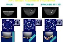
To test the hybrid system's 3D imaging capabilities, the Duke developers took a breast phantom and imaged it in three different orbits. In the first orbit -- in vertical axis-of-rotation (VAOR) -- the detector moves in a circular path around the breast at a 0° tilt. The second trajectory uses a tilted preal beam (TPB), where the detector is at a fixed 45° angle. In the third trajectory, the projsine orbit is where the camera moves in a semicircle range at a tilt of 15° to 45° (see images below).
 |
| Click here to enlarge this image. |
| All images courtesy of Duke University Medical Center Multi-Modality Imaging Lab. |
"The advantage of having the camera tilt is that we can now see more fully into the breast and closer to the chest wall," Madhav noted. "However, using orbits, such as TPB, insufficiently samples the breast, which causes shape distortion. Therefore, complex orbits with projsine improves sampling and restores the shape of the object."
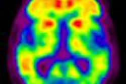
In their first patient study, the researchers imaged a postmenopausal 55-year-old woman with biopsy-proven adenocarcinoma, with ductal and lobular invasive features. She was injected with 660 MBq of Tc-99m sestamibi and the data was acquired at a 45° tilted orbit.
 |
| A postmenopausal 55-year-old woman with biopsy-proven adenocarcinoma, with ductal and lobular invasive features. |
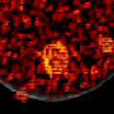
Duke researchers found what Madhav described as a "hot spot about 2 cm in diameter," which had been confirmed in a breast MRI scan earlier that day (see image above).
There are two items of note from the exam. "We see these streak artifacts, which we believe are due to the cardiac and HepTide uptake of the tracer in the body," Madhav said. "We also see a shape distortion, which is mostly due to an insufficient sampling of the orbit."
The Duke MMIL also is developing a prototype of a hybrid bed for acquiring images in patient studies, which the team plans to conduct before the end of the year.
 |
| The bed is made of 1/8-inch stainless steel, with angled sides for patient support and added strength for the cantilevered bed. |
An octagonal trough also allows for the system to rotate underneath the bed. A removable insert allows for radiolucent materials and imaging through the chest wall.
One drawback to the bed at this point is the inability to lower the bed more into the field-of-view, due to the physical constraints of the CT tube and its large width.
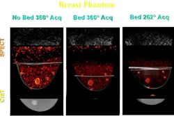
To determine how far the SPECT/CT system could see into the breast with and without the assistance of the bed, researchers took a 1,060-mL breast phantom and filled it with water and radioactivity, as well as a 1.8-cm lesion filled with radioactivity and CT contrast agent. Four external fiducial markers were placed around the breast phantom.
Three different views of the breast were taken. The breast was placed in the field-of-view without the bed, and later on the bed, while the team performed a 360° acquisition and a partial acquisition. The partial acquisition was done because the bed could be lowered further to see more breast volume.
For all three images, researchers used a projsine orbit for the SPECT system and a fixed tilt at 6.2° for the CT scan (see images below).

"Using the projsine orbit, we can see more to the chest wall. For the CT system, when we have the breast placed on the bed, as expected, we see less of the breast volume," Madhav said. "However, when we do a partial acquisition, we can see more into the chest wall."
With its research to-date, the Duke MMIL believes its dedicated dual-modality SPECT/CT system will be able to provide 3D volumetric breast imaging with functional and anatomical information. In addition, the fused images will provide more information of clinical value than independent systems alone.
By Wayne Forrest
AuntMinnie.com staff writer
July 18, 2007
Related Reading
Israeli group test-drives 64-slice SPECT/CT system for cardiac imaging, June 6, 2007
SPECT/CT offers on-target diagnosis in noncancerous bone disease, March 23, 2007
SPECT/CT improves sentinel node detection in overweight breast cancer patients, February 14, 2007
SPECT/CT enhances octreotide imaging, January 12, 2007
SPECT/CT implementation: More than plug and play, June 7, 2006
Copyright © 2007 AuntMinnie.com