On T1-weighted MRI, endometrial cysts generally show high signal intensity. Unfortunately, so does other gynecological pathology, such as corpus luteum cysts. Differentiating endometrial cysts from other hemorrhage adnexal cysts is important for adequate treatment, said Dr. Mayumi Takeuchi. Her group from the University of Tokushima in Tokushima, Japan, compared the MR manifestations of endometrial cysts with both 1.5- and 3-tesla MRI.
"Endometriosis is a common disease in women of reproductive age," Takeuchi said in a talk at the 2007 International Society for Magnetic Resonance in Medicine (ISMRM) in Berlin. "Recurrent hemorrhaging and repeated rupture may cause pelvic adhesions with infertility."
For this four-month long study, the authors included 10 patients with surgically proven cysts. Fast spin-echo T2-weighted images were obtained for all patients with both 1.5- and 3-tesla scanners (Signa Excited HD; Signa 3T HD, GE Healthcare of Chalfont St. Giles, U.K.). Five patients also underwent susceptibility-weighted imaging (SWI).
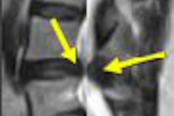
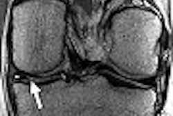
The shading sign, caused by the T2 shortening in an adnexal cyst, is an MR feature of endometriosis and is helpful for making the differential diagnosis, Takeuchi explained. In addition, the hemosiderin-laden macrophage deposits in the cyst wall are another pathologic feature of endometrial cysts, which results in signal voids on SWI.
"Because susceptibility-induced signal intensity loss may increase from 1.5T to 3T, the shading sign on T2-weighted images and signal voids due to hemosiderin deposition on SWI may (be) well visualized on 3T," Takeuchi and colleagues wrote in their ISMRM abstract.
For data analysis, cyst-to-muscle signal intensity ratio (SIR) was calculated based on region of measurements, and the SIR was defined as the cyst's signal intensity divided by the signal intensity of the piriform muscle, or the iliopsoas muscle, in the same plane.
According to the results, signal intensity of the cysts was lower at 3-tesla MRI than at 1.5-tesla based on visual inspection. The cyst-to-muscle SIR on 3-tesla imaging was 2.04 ± 0.90 versus 4.72 ± 2.34 on 1.5-tesla MR. The shading sign was seen on 3-tesla MR, but not on 1.5-tesla MR, in two lesions.
 |
| Endometrial cyst with the shading sign on T2-weighted 3-tesla MRI (above), which was not demonstrated on T2-weighted 1.5-tesla MR (below). Images courtesy of Dr. Mayumi Takeuchi. |
 |
In addition, signal voids caused by hemosiderin deposition in the cyst were seen more readily on 3-tesla SWI than on 1.5-tesla MRI. Finally, in an e-mail to AuntMinnie.com, Takeuchi said that using 3-tesla MRI did cut down on the exam time (eight minutes and 24 seconds versus nine minutes and 47 seconds at 1.5-tesla).
However, the group did acknowledge that susceptibility artifacts generated by air in the intestines were more prominent on 3-tesla MRI. "These results may reflect the higher sensitivity of 3T to the susceptibility-induced T2-shortening effect caused by intracystic, old hemorrhagic contents," the authors explained.
Additional research -- including the correlation of imaging exams with surgical or pathological results -- is needed to assess the value of 3-tesla MR in this patient population. Takeuchi said that her group plans to continue their investigation.
By Shalmali Pal
AuntMinnie.com staff writer
June 8, 2007
Related Reading
Age at endometriosis diagnosis is linked to breast cancer risk, April 12, 2007
Endometriosis does not increase long-term fracture risk, January 2, 2007
Copyright © 2007 AuntMinnie.com