In its new guideline for breast MR, the American Cancer Society (ACS) recommends against the routine use of the modality in women with ductal carcinoma in situ (DCIS). They cited figures from a Dutch study in which mammography outperformed MRI for detecting DCIS, with the former turning in a specificity of 95% (79.5% sensitivity) versus 90% (33.3% sensitivity) for the latter (New England Journal of Medicine, July 29, 2004, Vol. 351:5, pp. 427-437).
The results of a recent study jibe with the ACS' assessment: The Italian authors said that MRI could not be used as a diagnostic tool for evaluating microcalcifications, but that it did have other uses in DCIS. But first, Dr. Philippe Sébag from the Centre de Sénologie in Nice, France, outlined what mammography continues to offer in DCIS.
Microcalcifications on mammo
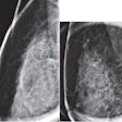
Mammography remains the gold standard for detecting DCIS, with a sensitivity that falls between 80% and 85%, Sébag said in a presentation at the 2007 Breast Course in Key Largo, FL. So what exactly do imaging experts look for on these mammograms?
"It's interesting to understand what happens (in DCIS) from a pathologic point of view," explained Sébag. "The neoplastic cell population is supported by connective tissue called the stroma. The stroma provides support and nutrition to the neoplastic cell, and that's the sign that we have to look for on mammography."
The presence of a major stromal reaction is related to invasive carcinoma and is frequently associated with microcalcifications. The latter serves as the most important mammographic feature in DCIS, Sébag said, adding that craniocaudal and mediolateral views were the best for microcalcification localization and characterization.
Most microcalcifications are smaller than 0.5 mm and are visible on mammograms with more than 100 micrometer resolution, Sébag stated. In 70% of the cases, the calcifications are in a ductal distribution and 90% of DCIS clusters have more than 10 flecks of calcification.
In addition, soft-tissue anomalies are associated with microcalcifications in women older than 70 years of age. Mammographic signs include masses, architectural distortion, or asymmetric density.
In the course syllabus ("The Breast Book 2007"), Sébag listed the morphological signs of DCIS from the most common to least common:
- Rod calcifications
- Branching-shape calcifications
- Punctate calcifications
- Predominantly punctate calcifications
With regard to invasive disease and high-grade DCIS, more than 40 calcifications on mammography means a 48% risk of occult invasive, which drops to 15% when there's less than 40 calcifications. Sébag pointed out that about one-third of malignant calcification clusters contain an invasive focus and that half of DCIS recurrences are invasive disease.
Keeping an eye on DCIS recurrence is particularly important in light of research out of France. Investigators at the Institute Bergonie in Bordeaux published the results of a retrospective study of bilateral ductal carcinoma in situ (DCIS) and found a local relapse rate of 20% in their patient population.
Moreover, this relapse took place in the contralateral breast and was invariably invasive cancer. The group warned that even after mastectomy, patients with DCIS required careful follow-up (Journal de gynécologie, obstétrique et biologie de la reproduction, March 19, 2007).
Microcalcifications on MR
For their investigation, Dr. Chiara Iacconi and colleagues selected 55 patients with mammographically found microcalcifications classified as BI-RADS 3-5. Iacconi's group is from University of Pisa and the Azienda Ospedaliero-Universitaria Pisana, both in Pisa.
Contrast-enhanced MR imaging was done on one of two 1.5-tesla units (Symphony, Siemens Medical Solutions, Malvern, PA; GE Healthcare, Chalfont St. Giles, U.K.). The protocol included a transverse T1-weighted localizing sequences and a sagittal fat-saturated T2-weighted sequence. Postprocessing included maximum intensity projection (MIP) reconstruction and time-intensity curves in the region of interest (ROI).
Enhancement morphology was divided into four categories:
- Absent enhancement
- Dendritic enhancement
- Nodular enhancement
- Dendritic and nodular enhancement
According to the results, microhistology identified 26 malignant lesions and 29 benign lesions. In the 26 cancers, enhancement morphology was dendritic and nodular enhancement in the majority of cases (14). No statistically significant difference was found between mammography and MRI in the diagnosis of malignant versus benign lesions, the authors stated.
Of the 55 patients, 48 had contrast enhancement on MRI and seven had no enhancement, with one of six patients having DCIS. The group tallied the following results for sensitivity, specificity, positive predictive value (PPV), negative predictive value (NPV), and accuracy:
|
"In our study, the sensitivity of contrast-enhanced MRI … was higher … however, the false-negative (n = 7) and false-positive (n = 7) MRIs strengthen the concept that mammography is the technique of choice for detecting microcalcifications and that stereotactic biopsy is mandatory for characterizing BI-RADS 3-5 microcalcifications," the authors wrote (La Radiologia Medica, March 2007, Vol. 112:2, pp. 272-286).
However, their data showed that MRI did perform better than mammography for the assessment of disease extent and was better for evaluating breast quadrant involvement, they added. In addition, in 15 of the 26 patients with malignancies, surgical treatment was planned on the basis of MRI, not mammography, findings.
Iacconi's group concluded that MRI's true role in DCIS could be as a preoperative tool for evaluating lesion size, multifocality, and multicentricity in breast-conserving surgical candidates.
By Shalmali Pal
AuntMinnie.com staff writer
April 20, 2007
Related Reading
Spiral MRI technique picks up what mammo misses in DCIS, March 15, 2007
Molecular markers: Putting too fine a point on DCIS? March 15, 2007
Breast pathology lacks standardization, particularly in DCIS, March 13, 2007
Breast MR shows exceptional sensitivity for spotting DCIS, January 12, 2007
Copyright © 2007 AuntMinnie.com


