KEY LARGO, FL - Superior pathology results can directly impact the treatment of breast cancer and its subsequent outcomes. Part and parcel of good pathologic results is the correlation of pathology with mammography, especially in cases of ductal carcinoma in situ (DCIS). Unfortunately, pathology practice is not always done at the most optimal level, according to a speaker at the Breast Course 2007.
"The pathologist's ability to establish diagnosis is … compromised when imaging studies are unavailable," said Dr. Michael Lagios in a talk he gave on Monday. DCIS is one area where subpar pathology results can have serious consequences, he added.
"We all recognize that tumor size is critical for treatment decisions," said Lagios, who is the medical director of Breast Cancer Consultation Services in Tiburon, CA, and is a clinical associate professor in pathology at Stanford University in Stanford, CA. "The best estimate of tumor size results from correlating imaging and histologic data, but this is something that is often neglected."
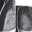
To achieve even "adequate" mammographic and pathologic correlation, the following elements must fall into place: preoperative images, localization studies, specimen radiographs, and postsurgical mammograms. Ideally, this will happen intraoperatively, Lagios said, because correlating results retrospectively is difficult.
But that kind of synchronicity between different medical specialties is not easy to achieve, Lagios conceded. Furthermore, the practice of pathology is often treated as "uniform and reproducible," when it is actually quite variable, he added.
Lagios said that there are two minimal requirements for the pathologic study of breast resection with DCIS: the aforementioned mammographic-pathologic correlation and the complete and thorough examination of the resected tissue.
In terms of the latter, misclassification can occur when the pathologist relies on gross examination ("eyeball estimates") of the specimen or engages in random tissue sampling, which can mean missed invasive foci, he said.
"Eyeball estimates are very common. Many of my colleagues will measure a breast cancer by holding the specimen in the left hand and the ruler in the right and the two will never meet," he said.
In addition, defining the margin status pathologically is important and cannot be based on less than complete tissue processing (CTP), he emphasized, noting that mammographically detected DCIS is nonpalpable and microscopic (between 5 mm and 10 mm).
Assessing tumor size is another area where common shortcuts can lead to long-term problems, such as inappropriate disease treatment. "Pathologists have been instructed from their infancy to section things, not to achieve the largest diameter of the tumor but to 'bread loaf' it. Depending on their awareness of the pre-op imaging or what section they take, they might get any number of sizes but many of them will be much smaller than the actual tumor size," Lagios explained.
He recommended that DCIS specimens should either be limited to a single slide or laid out on multiple slides that have a known sequential relationship to each other, as well as a known block thickness.
While some may argue that (CTP) is too time-consuming and expensive for widespread use, Lagios countered that studies have shown that a rigid CTP protocol can identify patients with a low risk of recurrence, as well as women who would not benefit from radiation therapy or adjuvant treatment (New England Journal of Medicine, May 13, 1999, Vol. 340:19, pp. 1455-1461; Cancer, February 15, 1989, Vol. 63:4, pp. 618-624).
Lagios' call for a team approach to breast cancer reflects the growing connection between the medical specialties. In an essay featured in the Breast Course syllabus, Barbara Rabinowitz, Ph.D., pointed out that "the roles of many breast care providers have evolved dramatically over the last two decades…. For example, hormone assays and sentinel node biopsy have expanded the role of pathologists and radiologists, as information about node involvement and disease metastases … now helps to guide many … therapy decisions" ("The Breast Book 2007").
This convergence illustrates a shift that must take place in medicine from multidisciplinary care to interdisciplinary care, according to Dr. Robert Kuske, who chaired the session.
Kuske explained that in the current multidisciplinary model of healthcare, a woman with breast cancer would be shuttled between various doctors, never really knowing if those specialists are communicating with each other. This setup is convenient for the physicians, but not for the patients, Kuske said.
In an interdisciplinary model, the patient would be treated at a single center with each doctor coming to her, then consulting with each other throughout the diagnostic and treatment process, he added.
By Shalmali Pal
AuntMinnie.com staff writer
March 13, 2007
Related Reading
HIFU breast cancer treatment may leave immunity-building antigens, February 26, 2007
Breast MR shows exceptional sensitivity for spotting DCIS, January 12, 2007
Breast MRI measures biologic significance of DCIS, October 18, 2006
Copyright © 2007 AuntMinnie.com
