CT start-up firm Koning of West Henrietta, NY, has launched its first conebeam CT system, Koning CT for Breast and Extremities, at this week's RSNA show in Chicago.
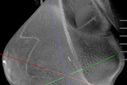
The device uses a cone-shaped x-ray beam and a digital flat-panel detector to capture volumetric data, and produces high-resolution 3D images with true isotopic resolution, according to Koning. It also reduces the patient dose as compared to current CT devices, the company said: A pilot study at the University of Rochester Medical Center found that Koning CT for Breast and Extremities showed potential to outperform mammography with better image quality and diagnostic information at radiation levels comparable to a two-view mammogram.
The device consists of a cushioned exam table with a cutout in the center, as well as a horizontal x-ray assembly. Patients lie down and the breast to be imaged is suspended through the opening while images are taken in a 360° circle in 10 seconds. No breast compression is necessary. Volumetric images are displayed in multislice, multiplanar format as well as 3D.
The company has begun preparing data to submit Koning CT for Breast and Extremities to the U.S. Food and Drug Administration for clearance, which it hopes to receive in 2007.
By AuntMinnie.com staff writers
November 26, 2006
Related Reading
Mercury, Koning combine for conebeam CT, November 2, 2006
Road to RSNA, Koning, October 26, 2006
Imaging start-up to install conebeam CT breast imaging prototype, May 1, 2006
Breast CT developers aim to 'revolutionize' mammography, December 20, 2005
Researchers make inroads with breast CT, gear up for clinical test, July 25, 2005
Copyright © 2006 AuntMinnie.com