A computer-aided diagnosis (CAD) multimodality workstation can improve the performance of radiologists in differentiating malignant and benign breast lesions on mammography and sonography, according to research published in the August issue of Radiology.
"We have shown that the use of our multimodality intelligent workstation improved the performance of both breast radiologists and breast imaging fellows in the task of differentiating malignant from benign breast lesions on mammograms and sonograms," wrote a research team from the University of Chicago in Illinois (Radiology, August 2006, Vol. 240:2, pp. 357-368).
The study team led by Karla Horsch, Ph.D., performed a retrospective review of 97 breast lesions from a multimodality database. Five breast radiologists and five breast imaging fellows gave confidence ratings and patient management decisions, both with and without computer aid. The lesions were unknown to both the observers and the computer.
The institution utilized a multimodality workstation that displays four-on-one breast mammograms, with regions of interest containing a given lesion from the standard and special mammographic views, and the sonographic views. The mammographic and sonographic computer output is presented in three different ways, according to the researchers.
First, separate estimates of the probability of malignancy (PM) are calculated for a given lesion's mammographic and sonographic views; also, a computer analysis is presented pictorially through a color-coded display of images automatically selected from either mammographic or sonographic reference libraries. In addition, computer analysis is presented graphically, with the lesion's mammographic or sonographic PM or feature value shown relative to the histogram of malignant and benign lesions from the appropriate reference library, according to the researchers.
Use of the CAD workstation improved of the average performance of the 10 observers, with a statistically significant increase in Az value (0.87 to 0.92; p < 0.001), partial Az value (0.47 to 0.68; p < 0.001), and sensitivity (88% to 93%; p = 0.005). A statistically significant difference was not found in the specificity without and with the computer aid (66% to 69%; p = 0.20).
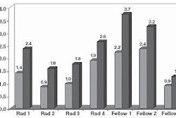
All observers changed the patient management decision for an average of 2.5 malignant lesions from follow-up to biopsy and for 0.6 malignant lesions from biopsy to follow-up. All observers changed the patient management decision for 2.0 benign solid lesions from follow-up to biopsy and for 3.3 benign solid lesions from biopsy to follow-up, according to the study team.
In other findings, the computer aid resulted in at least one observer correctly changing the management decision from biopsy to follow-up for 24 of the benign lesions and incorrectly changing it from follow-up to biopsy for 14 of the benign lesions, according to the authors.
The researchers acknowledged limitations of the study, including the lack of inclusion of any lesions that had not undergone biopsy or aspiration in their clinical evaluation. This might have affected the sensitivity and specificity of the radiologist user, they stated.
In addition, the study findings can't be used to determine whether the radiologists' performance with the multimodality workstation were significantly improved in comparison with the mammographic (single modality) workstation, according to the researchers.
Both breast imaging radiologists and breast imaging fellows showed statistically significant improvement in Az and partial Az values, the authors concluded.
"In addition, the sensitivity of the breast imaging fellows also improved at a statistically significant level," the authors wrote. "Such multimodality intelligent workstations, therefore, hold promise for the improvement of the performance of radiologists in the task of differentiating malignant and benign breast lesions on mammograms and sonograms, and transition and further validation of multimodality workstations in the clinical arena are warranted."
By Erik L. Ridley
AuntMinnie.com staff writer
August 17, 2006
Related Reading
Pilot study: Entropy-driven CAD zips through vast breast image database, August 2, 2006
CAD interval change analysis boosts mammogram accuracy, July 7, 2006
Breast CAD takes aim at architectural distortion, June 30, 2006
Breast CAD put to the test in high-volume community practice, June 26, 2006
CAD finds breast cancers that radiologists missed for years, March 6, 2006
Copyright © 2006 AuntMinnie.com