Breast CT has long been dismissed as a breast imaging option due to its higher radiation doses relative to mammography. But if the dose issue could be resolved, CT might just have some potential, according to imaging start-up Koning of Rochester, NY.
Koning's team believes that a new kind of CT, conebeam computed tomography (CBCT), could revolutionize breast imaging by producing fast, efficient, and detailed scans, not only saving lives by finding cancer more quickly, but alleviating the discomfort of breast compression and reducing the inevitable anxiety of waiting for x-ray results. The company plans to install its first clinical unit at the University of Rochester Medical Center (URMC) in New York in May.
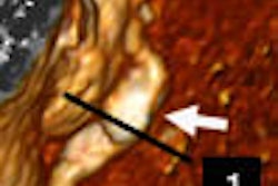
The device, called Koning CT, produces individual slices as well as 3D images of breast tissue, whereas mammography produces 2D images with structures superimposed on top of each other. Such superimposition can often hide small masses or tumors, according to the company's CEO and chief operating officer, John Neugebauer.
"Digital mammography has the same limitation when it comes to overlapping layers of tissue that can hide small tumors," Neugebauer said. "New tomosynthesis units which may be on the market soon will address some of the problem. But since Koning CT has full 360° capture and 3D reconstruction, it will routinely image small tumors and masses in the 2-3 mm range."
Koning was established as a technology spin-off in 2002 by Ruola Ning, Ph.D., of URMC's department of radiology. A biomedical engineer, Ning has been researching conebeam technology for more than 20 years, and Koning has won almost $11 million in grants from the National Institutes of Health (NIH), including a 2005 Small Business Innovation Research (SBIR) award of $2.65 million to develop conebeam CT for commercialization.
The company is working with Excell Partners of Canandaigua, NY, and URMC's Office of Technology Transfer to bring CBCT to market. URMC has put down $300,000 for CBCT patents, and has licensed the technology to Koning to make, use, and sell.
Koning CT's configuration includes a table designed to allow the patient to lie prone, the breast hanging but stabilized, rather than compressed. The x-ray source sends cone-shaped beams to a flat-panel digital detector, positioned under the table, which rotates around the breast a full 360º in 10 seconds to capture a full set of data volumetrically.
Patented software produces isotropic 3D images of the breast: Each image slice taken from any planar orientation will exhibit equal resolution in the x, y, and z axes, so that there is less chance of missed tumors; the device's spatial resolution for these axes is 0.1 mm to 0.5 mm, as compared to 3 mm for single-slice CT and 1 mm for spiral multislice CT, according to Koning.
The table and the position of the breast make Koning CT a more comfortable alternative to mammography, according to the company, and the radiation dose is no higher than the dose for a standard two-view mammography exam.
President and chief technology officer Ning chose to focus on breast imaging first because of conebeam's potential to improve on what he sees as the limitations of current methods. Also, the current generation of flat-panel digital detector technology, while not yet ready for whole-body CT use, is now capable of capturing volumetric data for limited field-of-view applications, such as breast, hands, feet, and extremities, Neugebauer said.
"As flat-panel detector technology further matures, CBCT will become smaller, cheaper, and faster, because it doesn't require multiple arrays or motorized tables to scan the patient," he said. "We think that within 15 years conebeam will be widely used, even for whole-body applications."
Koning's researchers are not the only ones racing to explore CBCT. In addition to big multimodality OEMs, researchers from the University of California are throwing their hats into the CBCT ring. The UC group includes radiology professor John Boone, Ph.D.; Drs. Karen Lindfors and J. Anthony Seibert from Davis; and Dr. Thomas Nelson from San Diego. They presented preliminary clinical results at the 2005 RSNA meeting in Chicago taken from a prototype device they have developed that was funded by $6 million in grants from the California Breast Cancer Research Program, the National Cancer Institute, and the National Institute for Biomedical Imaging and Bioengineering.
Koning carries eight patents and has four pending that cover the technology's method apparatus and the breast imaging application, which it hopes will help the firm maintain a place in a market dominated by large multimodality vendors, Neugebauer said.
Koning's greatest challenge now is to prove the technology. The company is in discussion with the U.S. Food and Drug Administration about the appropriate clearance process for its device, which it is developing as a diagnostic, rather than a screening tool. Koning plans to highlight CBCT at this year's RSNA meeting, and hopes to place several units in 2007.
"The actual science and technology are well in place," Neugebauer said. "Our efforts over the next six months will be focused on demonstrating the device's performance."
By Kate Madden Yee
AuntMinnie.com contributing writer
May 1, 2006
Related Reading
Breast CT developers aim to 'revolutionize' mammography, December 20, 2005
Researchers make inroads with breast CT, gear up for clinical test, July 25, 2005
Copyright © 2006 AuntMinnie.com