Everyone knows that breast MRI has high sensitivity but poor specificity. The opposite holds true for whole-body PET imaging in breast cancer, which has high specificity but lacks sensitivity. The better molecular imaging modality may be positron emission mammography (PEM), especially for recognizing primary breast cancer.
"Because the spatial distance between the detector plates is significantly less (in PEM) than for whole-body PET, the resolution capacity is higher," explained Dr. Lorraine Tafra from the Breast Center in Annapolis, MD. Tafra's pilot study showed that PEM was particularly adept at spotting ductal carcinoma in situ (American Journal of Surgery, October 2005, Vol. 190:4, pp. 628-632).
Tafra's co-authors are from various institutions in Metairie, LA; Lutherville, MD; Winston-Salem, NC; and Philadelphia. Deepa Narayanan of Naviscan PET Systems in Rockville, MD, is also a contributor.
The team collected data from 44 women, the majority of whom were postmenopausal with a median age of 57 years. The women underwent PEM with a median dose of 13 mCi FDG. The median compression breast thickness was 63 mm.
Two readers assessed whether PEM could detect the primary lesion, find multifocal disease, detect incidental cancers, and predict margin status. The PEM results were not used to guide surgical therapy, but they were compared to surgical pathology outcomes.
The median tumor size of the primary lesion was 22 mm on pathology, 22 mm on PEM, and 20 mm on mammography. The majority (70%) were nonpalpable. PEM correctly interpreted the primary lesions as probably malignant for 89% of the patients.
Three lesions were not found by PEM, including a 6-mm low-grade tubular carcinoma and a 1-mm breast lymphoma. Variables in the metabolic activity of breast cancer may have been the reason that PEM failed in these cases, the authors suggested.
PEM found 10 incidental breast abnormalities, of which five were malignant; PEM determined four out of the five. The modality correctly predicted multifocal disease in 64% of the women, and it accurately predicted positive margin status in 75% of the women undergoing partial mastectomy.
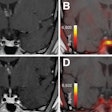
Finally, PEM identified a small, suspicious abnormality that turned out to be ductal carcinoma in situ (DCIS), although it was benign on mammography. "PEM appears promising in the depiction of DCIS. The presence of DCIS can be an enormous surgical challenge," the authors stated. "Unless the DCIS is associated with mammographically detectable features such as microcalcifications, it is very difficult to characterize the extent of disease." In addition, all of these women had dense breasts, which PEM was able to penetrate.
Had the PEM information been available to the surgeons, it could have offered guidance for more definitive surgery and negated the need for re-excision, the authors concluded, calling for further studies on the negative and positive predictive value of PEM, as well as how it compares to other modalities such as breast MR.
Tafra and other co-authors, including Dr. Wendie Berg, Ph.D., from the American College of Radiology Imaging Network (ACRIN) in Lutherville, MD, will update the use of PEM at the 2005 RSNA meeting in Chicago. Their scientific paper will discuss standard uptake value (SUV) differences in breast epithelial subtypes on PEM (SST01-09).
By Shalmali Pal
AuntMinnie.com staff writer
November 10, 2005
Related Reading
PET mammography unit shows promise for DCIS, June 22, 2005
PEM development picks up pace, February 7, 2005
Copyright © 2005 AuntMinnie.com


