Dual-energy CT may find itself forging a new path in virtual colonoscopy by not only finding colorectal lesions but characterizing them in ways other noninvasive techniques cannot, according to a presentation at the International Society for Computed Tomography (ISCT) conference.
For patients at higher risk of harboring polyps with advanced histology or colon cancer, the use of dual-energy VC (also known as CT colonography or CTC) with contrast could help highlight high-risk lesions while allowing patients who probably have lesions with benign histologies to be managed more conservatively -- thus ensuring that intervention is reserved for patients who need it.
Dual-energy imaging -- which helps find tumors by highlighting the iodine they absorb following contrast media administration -- isn't a new invention; in fact, it's been around in one form or other since the 1970s. But dual-energy CT has gotten a big boost in recent years with the advent of dual-source CT scanners, which employ dual sets of x-ray tubes and detector arrays that can scan at two different energies simultaneously.
These days, dual-energy imaging is indicated for a number of applications, including virtual noncontrast images of the liver and kidneys, bone removal, runoff and plaque imaging, renal stones and gout, lung perfusion, myocardial perfusion, and more.
But dual-energy VC? At this point it's for research only and really is just getting started, said Dr. Anno Graser from the University of Munich in Germany in his ISCT presentation. Even his 2009 article on the potential of dual-energy CT in the abdomen (European Radiology, January 2009, Vol. 19:1, pp. 13-23) specifically excluded its use for virtual colonoscopy due to technical limitations of the scanner.
"The limitations of the first-generation dual-source scanner, namely the limited size of the B-detector ... and also the noise issues on the low-kV tube with 80 kV, really prevented us from acquiring CT colonography images with dual energy," said Graser, who is an assistant professor of radiology.
But the limitations were resolved with the introduction last year of a second-generation dual-source scanner (Somatom Definition Flash, Siemens Healthcare, Erlangen, Germany), which enables dual-energy CTC datasets to be acquired at 100/140 kVp, Graser said.
Virtual colonoscopy, which has shown high accuracy in detecting clinically significant polyps 1 cm or larger in several large trials, is a noncontrast low-dose CT exam that relies on morphology, size, internal density, and mobility to characterize polyps.
Unfortunately, VC "gives no information about the histology of lesions," Graser said. When you detect a polyp using single-source VC, "you can't tell if it's going to be an adenoma or a hyperplastic polyp -- it's just not possible," he said.
But with the use of dual-energy CT and IV contrast "we can determine the contrast media uptake of polyps [iodine concentration measured in g/mL] and we can also look at baseline [HU] densities ... from the so-called virtual nonenhanced images," Graser said. "We can then directly measure enhancement in Hounsfield units."
This technique is not suitable for digital subtraction of tagged fluid from the colon, Graser said. Once the iodine is subtracted, one is left with nonenhanced residual fluid.
Nor is dual-energy CTC a low-dose exam suitable for screening CT colonography. It requires contrast and delivers a standard radiation dose, making it more for high-risk patients, he said. Patients with stenosing lesions resulting in incomplete colonoscopy constitute a sizeable percentage of the dual-energy CT virtual colonoscopy patients, Graser said.
Images are acquired in two positions, as in standard CTC: first, a single-source noncontrast prone scan is taken, followed by contrast-enhanced dual-energy supine imaging, he said.
 |
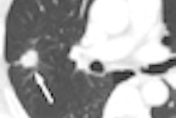
| Dual-energy virtual colonoscopy is acquired using a noncontrast prone exam followed by a contrast-enhanced supine scan on a dual-source scanner (Somatom Definition Flash). All images courtesy of Dr. Anno Graser. |
"Potential indications for this technique include examination of the entire colon in patients with stenosing carcinoma in order to search for synchronous polyps and cancer," Graser said. "It can also be a true one-stop examination ... for local tumor and nodal staging, as well as assessment for metastases."
It can also be used to evaluate benign strictures and to rule out recurrence at the site of anastomosis after colorectal cancer resection, he added.
 |
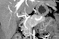
| CTC found several significant lesions in this patient, including a sessile polyp in the sigmoid colon, and another in the fold of the hepatic flexure, suggesting polyposis of the colon. Translucency rendering of polyp in the hepatic flexure, color coded red, indicates soft-tissue density. Below, dual-energy CT of the lesion is orange in color (determined to be 52 HU per comparison with virtual noncontrast image) and demonstrates contrast enhancement for the lesion as well as a lymph node. The patient was diagnosed with B-cell lymphoma. |
 |
CT colonography is a new potential indication for dual-energy CT, one that has a unique ability to characterize colonic lesions based on contrast enhancement, Graser said.
"Dual-energy CT colonography is able to show high-resolution imaging of the distended large bowel," he said, "but you need to have a noncontrast and a contrast-enhanced exam to perform an evaluation of the entire colon."
By Eric Barnes
AuntMinnie.com staff writer
June 2, 2010
Related Reading
Small colon polyps on VC suggest presence of larger lesions, April 16, 2010
DSCT beats other methods for noninvasive gout diagnosis, March 30, 2010
Dual-energy VC makes tagged materials disappear, October 29, 2008
Five-modality Munich trial finds high sensitivity for OC, VC, November 12, 2007
Copyright © 2010 AuntMinnie.com