The latest version of a novel 3D virtual colonoscopy viewing method significantly shortened reading times compared to 2D in a new study, though sensitivity was unchanged from conventional methods, researchers from South Korea reported.
An earlier version of the software also produced shorter interpretation times and equivalent sensitivity in a smaller 2005 study reported by AuntMinnie.com.
The present research, from Seoul National University College of Medicine and Ewha Woman's University Hospital, both in Seoul, aimed to compare primary 2D data interpretation (with endoluminal 3D confirmation) to a novel 3D dissection technique.
Study authors Drs. Se Hyung Kim, Jeong Min Lee, Hyo Won Eun, and colleagues evaluated the two methods in terms of interpretation time and sensitivity for colonic polyp detection, with same-day colonoscopy and segmentally unblinded virtual colonoscopy results used as the reference standard.
Several investigators, including Pickhardt et al from the University of Wisconsin in Madison, have found encouraging performance with the use of 3D endoluminal interpretation in virtual colonoscopy (VC or CT colonography [CTC]) for interpretation of CTC datasets, and a "growing minority" of radiologists now prefer it, the researchers wrote.
"Nevertheless, the 3D approach, a virtual imitation of colonoscopy, has limitations in that it does not enable the inspection of 'blind spots,' such as the parts of the mucosa that are hidden by colon folds," they wrote. Even four fly-through passes of the colon don't eliminate these blind spots completely, although they increase interpretation time (Radiology, September 2007, Vol. 244:3, pp. 852-864).
In contrast, virtual dissection can prevent the blind spots created "when all of the projection rays are perpendicular to the centerline (eye point)," the authors explained. The colon dissection software used in the study (Perspective Filet View; Philips Medical Systems, Andover, MA) "not only unrolls the colon but also bends the projection rays from the eye point to show both sides of the folds and the areas between tight folds," they wrote. As a result, a single pass across the flattened colon enables visualization of the entire colonic mucosa.
The study examined 96 consecutive patients (56 men, 40 women; mean age, 54.8 years) who underwent colonoscopy and VC on the same day. The cases were the first of 200 datasets from colorectal cancer screening patients, following exclusion of 32 patients for prior colorectal surgery, family or personal history of polyps or cancer, positive fecal occult positive blood test, polyposis, or other signs of heightened risk for colorectal cancer.
 |
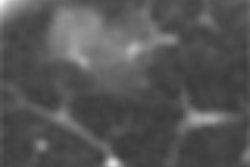
| Software-generated supine (above) and prone (below) 3D virtual dissection images. Each 10° area at the top and bottom of the image is added to the full 360° view of the colonic surface and displayed with a transparently shaded color so as not to miss any lesion. The colonic surface can be observed quickly by using the top scroll bars. Note that the polyp (arrows) remains unchanged on both images, but the lump of feces (arrowheads) changes in position. A 5-mm adenomatous polyp was confirmed at both colonoscopy and biopsy. Images (Fig. 1c) republished with permission of the authors and the Radiological Society of North America from Se Hyung Kim, M.D.; Jeong Min Lee, M.D.; Hyo Won Eun, M.D.; Min Woo Lee, M.D.; Joon Koo Han, M.D.; Jae Young Lee, M.D.; and Byung Ihn Choi, M.D.; "Two- versus Three-dimensional Colon Evaluation with Recently Developed Virtual Dissection Software for CT Colonography," Radiology 2007;244:852-864, © RSNA 2007. |
 |
All patients underwent cathartic bowel cleansing by ingesting 4 L of polyethylene glycol solution (Colyte-F; TaeJoon Pharmaceuticals, Seoul, South Korea) the day before scanning. Following colonic insufflation with room air to tolerance, patients were imaged on a 16-detector-row scanner (Sensation 16, Siemens Medical Solutions, Erlangen, Germany). Scanner settings included 16 x 0.75-mm collimation, 120 kVp, and 50 and 200 effective mAs for both prone and supine positions.
Real-time interpretation by two experienced radiologists consisted of a 2D examination with traditional 3D endoluminal software (Rapidia, Infinitt, Seoul, South Korea) for confirmation and problem solving.
The images were also interpreted using the 3D virtual dissection method that presented the data in an approximately 380° circular view, which included 360° plus a 20% overlap on each of the colons along an automatically calculated centerline.
 |
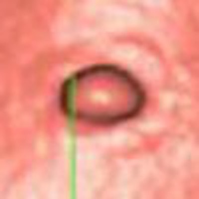
| Above, sessile 7-mm polyp in sigmoid colon of 60-year-old man. Virtual 3D views of supine (left) and prone (right) datasets show a small sessile polyp (arrow) in the sigmoid colon. Below, transverse 2D CT colonographic images of supine and prone (not shown) datasets also show the polyp (arrow) in the sigmoid colon. Bottom, conventional colonoscopic findings confirm the presence of the lesion (arrow). Biopsy was subsequently performed, and the final pathologic diagnosis was of an adenomatous polyp. Images courtesy of Dr. Se Hyung Kim. |
 |
 |
Two radiologists independently analyzed the CT data, both per polyp and per patient. The sensitivity of 2D and 3D were compared by using a McNemar test, and interpretation time was measured for each method, Kim and colleagues wrote.
Immediately following CT colonography, patients were sedated (midazolam, Bukwang Pharmaceutical, Seoul, South Korea) for optical colonoscopy performed by experienced gastroenterologists.
The final results of colonoscopy and unblinded CTC data revealed 134 polyps 2-30 mm in diameter in 55 of the 96 (57%) patients, and 99 polyps 5 mm or smaller. Of these, 23 were 6-9 mm in size, and 12 were 10 mm and larger. Seven of nine advanced lesions were tubulovillous adenomas, including a 15-mm and a 30-mm lesion, but no colorectal cancers were found.
Compared with the conventional 2D-3D polyp detection method, primary 3D interpretation using the filet view software showed "comparable per-polyp (77% and 69% for two readers) and per-patient (77% and 73% for two readers) sensitivities and comparable per-patient specificity (99% and 89% for two readers) for the detection of polyps 6 mm in diameter or larger," the authors wrote. Overall per-polyp and per-patient sensitivities did not differ significantly between the two interpretation methods for either reader (p > 0.05 for both).
However, interpretation times using the 3D virtual dissection software were significantly faster, completed in a median 9.4 minutes (range, 4.5-24.0 minutes) for reader 1 and 9.6 minutes (range, 4.3-26.7 minutes) for reader 2. Using the 2D-3D method, median interpretation time was 14.1 minutes (range, 5.8-35.5 minutes) for reader 1 and 14.4 minutes (range, 5.1-38.3 minutes) for reader 2, a significant difference for both readers (p < 0.05).
Use of 3D virtual dissection compared with primary 2D with endoluminal 3D confirmation "significantly shortens the interpretation time in the detection of colonic polyps," the authors concluded. "It allows supine and prone images to be displayed adjacent to each other in a flat form and thereby facilitates intuitive and direct perception."
The new method shortens interpretation time both by enabling radiologists to simultaneously interpret prone and supine datasets, and by eliminating the need for antegrade and retrograde navigation required for 3D endoluminal viewing of CTC data.
Likely owing to the "wet" polyethylene glycol prep, most colonic segments (98.5%) had little in the way of residual fecal matter, and per-patient specificity was in the high range, the authors wrote.
The average number of false-positive lesions was higher at 3D interpretation compared to 2D, they added. "The initial interpretation of CT colonographic data was performed by using the primary 2D interpretation method with a 3D problem-solving technique," they wrote. "Therefore, among the 26 and 35 FP (false-positive) polyps identified only on the 3D view by readers 1 and 2, respectively, and which were not subjected to segmental unblinding, we cannot be sure how many true-positive lesions were missed at both initial CT colonographic interpretation and colonoscopy. If we had used a more sensitive 3D virtual dissection view at the time of the initial interpretation, the sensitivity of the 3D view might have been higher. This means that the number of FP polyps detected on the 3D view might have been lower."
Both per-polyp and per-patient sensitivities were greater with 3D than with 2D interpretation, "even though the differences between the two methods were not significant for either reader," Kim and colleagues concluded.
By Eric Barnes
AuntMinnie.com staff writer
August 28, 2007
Related Reading
New developments improve VC -- and colonoscopy, April 6, 2007
Virtual dissection reading method finds the same polyps faster, December 12, 2006
VC software highlights colon's unseen areas, June 16, 2006
'Filet view' VC software slices reading time, October 6, 2005
VC visualization tool combines advanced features, August 17, 2006
Copyright © 2007 AuntMinnie.com