A new study from Sweden affirms the superiority of thinner CT collimation for detecting colonic polyps, even when constant radiation doses result in noisier images.
"In the design of low-dose CT sequences, there is always a trade-off between the radiation dose and spatial resolution properties of the image data," wrote Dr. Abdülvahit Özgün and colleagues in April's American Journal of Roentgenology. "Low-dose protocols typically involve high tube voltage, low tube current settings (calling for thicker slices), and a high pitch. Recent patient studies using a single or two different low-dose milliampere-second levels in CT colonography or simulating low doses by postexamination modification of imaging raw data indicate that despite reduction of image quality below the level of 30 mAs, polyp detection in CT colonography remains acceptable" (AJR, April 2005, Vol. 184:4, pp. 1181-1188).
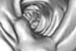
The research team, from Karolinska University Hospital in Stockholm, Sweden, built a latex phantom designed to be as close as possible to a real colon. A total of 240 synthetic polyps were handmade from the latex material and divided into three size groups: < 2 mm, < 2-5 mm, and < 5-10 mm.
"The polyps were evenly distributed among the groups, resulting in 80 polyps of each size interval," the authors wrote. "The shapes of polyps found in patients can be characterized as pedunculated; broad-based; ulcerated or depressed with elevated margins; or sessile or flat. Flat polyps were unique in that they were all rectangular with a size ratio width-height equal to or larger than 1. To simulate the in vivo situation as closely as possible, we manually manufactured 'polyps' of these four shapes. Their size and shape were visually confirmed with the help of optical magnification instruments and precision rulers."
The 24 colon phantoms, each containing 60 polyps (20 in each size group), were submerged in a water tank and scanned on a four-slice MDCT scanner (LightSpeed QX/i, GE Healthcare, Chalfont St. Giles, U.K.) using 12 protocols with various settings of slice thickness, pitch, and tube current. The tube potential was kept constant (120 kV) in all sequences.
"Estimates of the dose-length product (DLP) were calculated from measured normalized CT dose index-weighted (nCTDIw) values for the different beam collimations used -- that is, 5 (or 4 x 1.25), 10 (4 x 2.5), and 15 (4 x 3.75)," the group wrote. The standard setting, defined as a 100% dose level at a 30-mm scanning length, was 100 mAs, 1.5 pitch, 2.5-mm slice thickness, with dose calculations defined relative to the standard setting. A total of 45 data acquisitions were performed, including nine using a water-filled phantom and 36 without.
Three radiologists experienced in VC interpretation evaluated the images on a GE Advantage Windows 3.1 workstation using GE Navigator (version 2) software at a window width of 1,600 H, window level 400 H. Both 2D axial images and 3D endoluminal images were reviewed.
The results showed that at a constant dose, the polyp detection rate increased with increasing axial spatial resolution. Using the standard protocol (2.50-mm slice thickness, 1.5 pitch), the detection rate for all polyp sizes decreased from approximately 70% at 40 mAs to 55% at 40 mAs. For polyps larger than 5 mm, however, the detection rate remained nearly constant between 60-100 mAs tube current.
The researchers found a similar dependence on radiation dose for both 2D and 3D evaluation techniques, with detection improving as the dose rose. Image noise followed the predicted behavior closely, and varied linearly with the product of slice thickness and tube current, the group noted.
When the evaluation of 2D MPRs was divided into polyp size categories, the detection rate increased for larger polyps at all DLP levels. At 60% DLP relative to the standard protocol, for example, approximately 20% of the smallest polyps were detected. The detection rate rose to nearly 80% for medium-sized polyps and to 90% for the largest polyps.
"The axial spatial resolution -- that is, the slice thickness and pitch factor -- significantly affected the detection rate of polyps," the group wrote. "When the radiation dose (DLP) was kept constant and equal to that of the standard sequence ... the highest detection rate of polyps larger than 2 mm was obtained using the sequence with 1.25-mm slice thickness and 0.75 pitch, followed by the sequence with 1.25-mm slice thickness and 1.5 pitch (2D MPR technique). This is despite the significantly reduced tube current settings used with the narrower slice sequences to balance DLP (40 and 80 mAs, respectively)."
"The detection of polyps in the large bowel using a standard protocol can be improved without dose penalty by increasing the axial spatial resolution," the group concluded.
Still, thin-collimation scanning does take longer, and results in more images to store, the authors wrote. Also, they noted, the study results need to be validated in the latest scanner models with more detector rows.
"The results demonstrate that imaging for polyps larger than 5 mm can be performed at significantly reduced dose levels without compromising the detection rate," the group wrote.
By Eric Barnes
AuntMinnie.com staff writer
April 27, 2005
Related Reading
What virtual colonoscopy misses might not matter, August 18, 2004
Thinner collimation improves polyp detection -- sometimes, November 26, 2003
Thin slices, narrow collimation rule in virtual colonoscopy study, March 8, 2003
Copyright © 2005 AuntMinnie.com