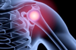
Ultrasound elastography can be a sensitive means of diagnosing rotator cuff tears in patients with painful shoulders, according to research from Alfa Scan Radiology Center in Giza, Egypt.
"Detection of tissue softening by elastography might predict tendonitis at an early stage before MRI, as the examination can be done, unlike MRI, guided by the pain location," said Dr. Naglaa Abdel Razek.
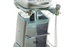
The research team sought to assess real-time ultrasound elastography for evaluating the supraspinatus tendon, studying 20 healthy volunteers and 40 patients presenting with shoulder pain. Elastography was performed using an EUB-7500 ultrasound system (Hitachi Medical, Tokyo) and electronic-array transducers of 7.5 and/or 13 MHz. Razek presented the research during a scientific session at the 2008 RSNA meeting in Chicago.
Tendon parts were evaluated by a semiquantitative score of different colors representing stiff tissue (blue) to softer tissue (green, yellow, and red).
In B-mode scanning, reviewers examined tendon insertion and the midportion and musculotendinous junction for tendon thickening, focal intratendinous abnormal high echogenicity, interruption of fibers, calcification, paratenonitis, and bursitis, according to Razek.
The elastography findings were then compared with the B-mode results, and for 20 patients, elastography was also compared with MRI findings. Arthroscopy was performed only when elastography was positive and MRI had a negative finding, Razek said.
In the 20 healthy volunteers, elastography showed blue color throughout the tendon, which is consistent with stiff normal tendon tissue and normal findings at grayscale, Razek said. In the patients with partial tears, elastography showed intratendinous color alterations (green, yellow, and red) not reaching the bursal or articular aspects. Patients with full tears showed color alterations reaching the bursal or articular surfaces.
The differences in tendon stiffness between the healthy volunteers and the patients were statistically significant (p < 0.0001). The elastography and MRI findings also showed good correlation (p < 0.001), Razek said.
In addition, elastography was able to diagnose tendinitis and mild synovial effusion in four cases (10%) that had false-negative findings on MRI. Elastography changed the diagnosis of partial tear into complete tear in two cases (5%), Razek said.
"The sensitivity and negative predictive value has been increased from 95% to 97% and from 87% to 93% by adding elastography to the conventional ultrasound technique," she said.
Elastography can also be used as an easy reproducible follow-up method to monitor treatment, Razek noted.
"Elastography is suggested as a complementary study to conventional high-resolution ultrasound for diagnosis of rotator cuff tendon tears in patients with painful shoulder," she concluded.
By Erik L. Ridley
AuntMinnie.com staff writer
January 15, 2009
Related Reading
Isotropic 3T MRI aids in diagnosing labrum, rotator cuff tears, December 29, 2008
MR arthrography outdoes 3-tesla MR on tendon tears, December 12, 2008
Mayo Clinic develops MR elastography for liver patients, November 19, 2008
Pictorial essay: 3D ultrasound reveals shoulder pathology, March 21, 2008
Study: Picking US over MRI for MSK imaging could save billions, March 6, 2008
Copyright © 2009 AuntMinnie.com