The American Academy of Family Physicians (AAFP) and the American College of Physicians (ACP) have jointly released new guidelines for diagnosing venous thromboembolism (VTE), including recommendations for utilizing ultrasound and other imaging studies.
In the first of four recommendations for clinicians, the Joint AAFP/ACP Physicians Panel on Deep Vein Thrombosis/Pulmonary Embolism stressed the use of validated clinical prediction rules to estimate pretest probability of venous thromboembolism. The authors also stated that in selected patients with low pretest probability of deep vein thrombosis (DVT) or pulmonary embolism, obtaining a high-sensitivity D-dimer assay is a reasonable option. If negative, that indicates a low likelihood of VTE, according to the panel (Annals of Family Medicine, January-February 2007, Vol. 5:1, pp. 57-62).
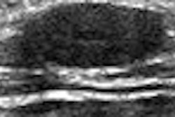
In the last two recommendations, the panel discussed the role of imaging. In patients with intermediate to high pretest probability of DVT in the lower extremities, they recommend using ultrasound.
"Use of ultrasound in diagnosing symptomatic thrombosis in the proximal veins of the lower limb is recommended for patients whose pretest probability of disease falls in the category of intermediate to high risk of DVT under the Wells prediction rule," the authors wrote. "Ultrasound is less sensitive in patients who have DVT limited to the calf; therefore, a negative ultrasound does not rule out DVT in these patients."
A repeat ultrasound or venography study may be required for patients who have suspected calf-vein DVT and a negative ultrasound, and for patients who have suspected proximal DVT and an ultrasound that is technically inadequate or equivocal, according to the authors.
"Contrast venography is still considered the definitive test to rule out the diagnosis of DVT," they wrote.
In the fourth and final recommendation, the organizations said that patients with intermediate or high pretest probability of pulmonary embolism require diagnostic imaging studies.
"Possible tests include ventilation-perfusion (V/Q) scan, multidetector helical computed axial tomography (CT), and pulmonary angiography," they wrote. "Recent systematic reviews indicate that CT alone may not be sufficiently sensitive to exclude pulmonary embolism in patients who have a high pretest probability of pulmonary embolism."
As for imaging's role in diagnosing venous thromboembolism, the authors concluded that there is strong evidence supporting the use of ultrasonography for diagnosing proximal DVT in symptomatic patients.
"Sensitivity is much lower in asymptomatic patients and for detecting calf vein DVT," the panel wrote. "Recent results suggest that newer CT technology for diagnosis of pulmonary embolism might have a higher sensitivity and specificity than seen in previous studies. In addition, it is likely that accuracy of CTs will improve with time as the technology evolves further."
By Erik L. Ridley
AuntMinnie.com staff writers
February 16, 2007
Related Reading
Guidelines released for diagnostic testing in suspected pulmonary embolism, December 6, 2006
CTA-CVT combo improves diagnosis of pulmonary embolism, June 1, 2006
Venous thromboembolism risk in colorectal cancer highest early after diagnosis, April 3, 2006
Diagnostic management of suspected pulmonary embolism often substandard, February 7, 2006
Model identifies low-risk patients with pulmonary embolism, January 24, 2006
Copyright © 2007 AuntMinnie.com