
Ultrasound technology has progressed to the point where low-cost, readily available 3D ultrasound techniques can compare to -- and compete with -- higher-cost techniques that are unavailable to many patients.
In our view, 3D ultrasound, a newer technique that is already well known and frequently used in obstetric diagnostics, has enormous value in other diagnostic areas, producing very useful data that can be compared with more sophisticated techniques such as CT and endoscopic retrograde cholangiography.
In our institution we have used 3D ultrasound in nonobstetric work for more than four years, and we have been able to compare our results with CT and cholangeographic findings in a large number of patients with pathology of the gallbladder and bile ducts.
All of the cases in this series were performed on a Voluson 530D scanner (Medison, Seoul, South Korea, now marketed in the Americas by GE Medical Systems of Waukesha, WI) equipped with a 3.5-MHz abdominal transducer, using a variety of techniques depending on the pathology found, to demonstrate the findings as clearly as possible.
The ultrasound features of the pathology were reviewed for the gallbladder and bile ducts seen in volumetric exploration, and were compared in some cases with other, traditional diagnostic imaging methods.
Part I: Gallbladder
Gallbladder adenomyomatosis
A 54-year-old man was sent to the ultrasound department for evaluation of digestive symptoms and pain in the right hypochondrium that had developed over several months. Findings were equivocal in the conventional 2D ultrasound exam with regard to a structure that did not seem to emit an acoustic shadow in the lower part of this organ, thus establishing a classic differential diagnosis between a non-pedunculated polypoid lesion and a hypoechoic calculus.
A volumetric scan was performed using a surface-rendering technique that confirmed a focal swelling of the gallbladder along its entire length where it was in contact with the liver. It was seen as an echogenic mass adjacent to the inferior liver, which confirmed the diagnosis of a gallbladder mass.
Following resection, histopathology yielded a diagnosis of gallbladder adenomyomatosis.
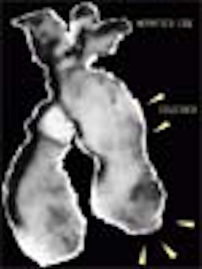
 |
| Figure 1. Volumetric reconstruction of the gallbladder using the surface-rendering technique. Swelling of the gallbladder wall is evident, as well as the existence of a polypoid lesion inferiorly (arrows). All images courtesy of Dr. O. Valls Pérez and Dr. M. E. Parilla Delgado. |
Gallbladder lithiasis and tumor
A 70-year-old woman with a long history of digestive problems, pain in the right hypochondium, and now a palpable mass in the same area, was sent to the ultrasound department for evaluation of this area.
Abdominal ultrasound showed the existence of a stone toward the neck of the gallbladder, and a solid tumorlike mass in the medial aspect of the organ measuring 3 cm in diameter.
The patient underwent surgery for suspected lithiasis and gallbladder tumor. Histopathology confirmed the existence of a stone and an adenocarcinoma.
 |
| Figure 2. Volumetric reconstruction of the gallbladder obtained with a surface-rendering technique. A tumor is clearly visible in the medial aspect, without signs of wall infiltration, and with a calculus toward the gallbladder neck. Note the fine, regular outline of the rest of the gallbladder wall. |
Cholecystitis with perivesicular abscess
Over the course of a week, a 69-year-old man with a history of gallstones had developed a fever of unknown origin and pain in the right hypochondrion.
An ultrasound study of the abdomen revealed a distended gallbladder containing microlithiasis, bile-plug syndrome, poorly defined internal walls containing a thick liquid material, and a poorly defined border with the adjacent liver bed.
The patient underwent emergency surgery, which confirmed the diagnosis of perforated cholecystitis with perivesicular abscess.
 |
| Figure 3. Volumetric reconstruction of the gallbladder obtained with a surface-rendering technique clearly depicts the distended gallbladder with microlithiasis, bile-plug syndrome, poorly defined internal walls containing a thick liquid material, and a poorly defined border with the adjacent liver bed. |
Emphysematous cholecystitis
A 64-year-old man with history of diabetes presented at the hospital with acute pain in the right hypochondrion. Physical examination revealed swelling in the area, and a gallbladder that was both palpable and extremely painful. The patient was sent to the ultrasound department for a study of the gallbladder region, which revealed an organ of normal size, without calculi and with very poorly defined walls.
A large perivesicular collection could be seen containing thick liquid and gas, as well as subhepatic and peritoneal fluid collections.
A diagnosis of emphysematous cholecystitis was made and confirmed at surgery. The patient progressed satisfactorily and was discharged a week later.
 |
| Figure 4. Volumetric reconstruction of the gallbladder using a surface-rendering technique and MediCut software to delineate and emphasize the pathology. Image shows the gallbladder and perivesicular fluid collection containing gas. |
Part II: Hepatic ducts
Caroli's syndrome
A 35-year-old woman with a history of recurrent jaundice that was intrepreted as viral hepatitis. In the most recent episode, the jaundice was accompanied by fever and general decline in her condition.
An abdominal ultrasound study confirmed the existence of multiple calculi within the intrahepatic ducts, which showed segmental enlargement alternating with areas of stenosis.
A diagnosis of Caroli's Syndrome was made, and confirmed by endoscopic retrograde cholioangiography and liver biopsy.
 |
| Figure 5. Volumetric reconstruction with combined surface-rendering and maximal transparency techniques shows numerous small calculi inside the enlarged hepatic and intrahepatic ducts, giving the liver a coarse appearance. |
Acute cholangitis
A 35-year-old man with a history of alcoholism presented at the hospital with jaundice, fever, epigastric pain and dilatation, and irregularity of the intrahepatic ducts, as well as poor general condition. The 3D ultrasound study confirmed the existence of dilatation and irregularity of the intrahepatic ducts, which were filled with a thick substance, alteration in the liver parenchyma, and diffuse enlargement of the pancreas.
Planar imaging showed the existence of multiple very superficial liver abscesses, evidence of acute cholioangitis, and the development of chronic pancreatitis aggravated by the consumption of alcohol.
The sonographic diagnosis was confirmed by laboratory results and by endoscopic retrograde cholangiography.
 |
| Figure 6a. Volumetric reconstruction using a surface-rendering technique enabled clear visualization of the complete dilated intrahepatic hepatic biliary tree, which demonstrated irregular contours and contained thick material. |
 |
| Figure 6b. Volumetric reconstruction with a surface-rendering technique shows one of the superficial abscesses seen in this patient. |
Part III: Bile ducts
3-D sonographic cholioangiography
The study of the biliary ducts with ultrasound brings enormous value when confronting jaundice syndrome, helping to define it and determine whether or not it is obstructive in origin.
It is possible to make the diagnosis using 2-D ultrasound, however, the images can be interpreted only by a physician who is highly trained in this technique. By first opacifying the bile ducts using contrast materials, 3D ultrasound images can be obtained (during the processing of volumetric reconstructions) that are comparable to those acquired with traditional invasive imaging methods such as percutaneous cholangiography, endoscopic retrograde cholangiography, etc.
A type of electronic opacification can also be obtained with this technique using MediCut image-processing software. This method can also be used to produce images comparable to those obtained with the traditional imaging methods mentioned above.
Three-dimensional ultrasound also helps to expand the field of vision, permitting the visualization of not only the bile ducts, but also the cause of the obstruction and the involvement of other organs.
All of the cases presented henceforth were produced using this method (inverted-range surface-rendering technique with MediCut). Also presented are other traditional imaging methods, which were used to confirm or verify the diagnosis.
Obstruction of the common bile duct by a tumor (adenocarcinoma) in the head of the pancreas
A previously healthy 60-year-old woman became gradually jaundiced over the course of a month, accompanied by itching and a general decline in health. She was admitted to the ultrasound department for an abdominal ultrasound to determine the cause of jaundice.
The examination detected a tumor in the head of the pancreas, and a volumetric image was reconstructed using an inverted-range surface-rendering technique and electronic "dissection" of the image.
 |
| Figure 7a. Inverted-range, surface-rendered volumetric reconstruction and MediCut processing clearly show dilatation of the intrahepatic ducts and gallbladder, along with marked distension of the common bile duct caused by tumor infiltration in the head of the pancreas. |
 |
| Figure 7b. Barium study of the stomach and duodenum confirmed widening of the duodenal loop, with straightening of the mucosal folds due to tumor growth in the head of the pancreas. |
Obstruction of the common bile duct and pancreatic duct by tumoral growth at head of pancreas
A 50-year-old man who developed a greenish jaundice over 15 days, along with general decline in health, was admitted to the ultrasound department for an abdominal scan, assessment of etiology of disease, and treatment. The study showed dilatation of the intrahepatic and extrahepatic bile ducts, with distal obstruction of the common bile duct, tumor at the head of the pancreas, and dilatation of the bile duct and pancreatic duct.
 |
| Volumentric reconstructions obtained with 3-D ultrasound (Figure 8a, above) are compared with images obtained using endoscopic retrograde cholangeography (Figure 8b, below). Note the similarity of information that both methods bring to the diagnosis, including tumor at the head of the pancreas, and dilatation of the bile duct and pancreatic duct. |
 |
By Dr. María Parilla Delgado and Dr. Orlando Valls Pérez
AuntMinnie.com contributing writers
May 5, 2003
English translation by Eric Barnes.
Drs. María E. Parilla Delgado (assistant professor of radiology) and Dr. Orlando Valls Pérez (professor of radiology) are from the department of imaging, Hospital Hermanos Ameijeiras, Havana, Cuba.
Related Reading
Cuban radiologists rely on transcranial ultrasound, July 4, 2002
Preoperative assessment of bile duct stones with sonography, November 7, 2002
Ultrasound differentiates causes of abdominal pain in kids, October 4, 2002
Ultrasound of the pancreas: what normal looks like, December 17, 2002
Dynamic clips aid US gallstone presentation, July 8, 2002