By using simple weight-based kilovoltage adjustments with already low current, significant radiation dose reduction and acceptable image quality can be achieved for pediatric CT exams, according to a study in the May issue of the American Journal of Roentgenology.
Researchers from Lucille Packard Children's Hospital at Stanford University in California also found that increased image noise was a fair trade-off for decreased dose and "no study was considered diagnostically compromised."
The lead author of the study is Dr. Jee-Eun Kim from Lucille Packard Children's Hospital and Gil Medical Center at Gachon University of Medicine and Science in Incheon, Korea (AJR, May 2010, Vol. 194:5, pp. 1188-1192).
Radiation dose from CT scans is a concern for young patients who potentially face increased risk of organ exposure to radiation over their lifetime. Until recently, the study noted, dose reduction efforts have focused on decreasing the current (mAs) and lowering kilovoltage (kVp) to decrease radiation exposure. However, the authors contended there has been little research on the effects and efficacy of those changes.
Retrospective analysis
The researchers retrospectively evaluated 120 children who underwent routine contrast-enhanced chest CT. Sixty pediatric patients were scanned in 2006 and 60 others were imaged in 2008. In both study groups, 30 consecutive children weighed less than 15 kg (33 lb) and 30 consecutive children weighed between 15 and 60 kg (33 and 132 lb).
All CT studies were performed on a 64-slice scanner with automatic tube current modulation (CARE Dose 4D, Siemens Healthcare, Erlangen, Germany). In 2006, the CT protocol called for 120 kVp, with a reference mAs of 65. In 2008, the protocol was 80 kVp for pediatric patients less than 15 kg and 100 kVp for patients who weighed 15 to 60 kg, with reference mAs reduced to 55.
Radiation dose to the patient was measured by two standard dose indicators -- CT dose index volume and dose-length product -- and calculated by the CT scanner for each study.
The study defined CT dose index volume as the weighted CT dose index divided by the pitch; it represents the average dose throughout a circular pediatric phantom (160-mm diameter). The dose-length product is related to energy imparted to organs and used to assess the overall radiation of a CT exam; it's equal to CT dose index volume and the length of the scan in centimeters.
Effective dose was estimated using the dose-length product method with previously published age-dependent conversion factors.
Radiation reduction
With the adjustments in weight-based kVp and adding decreased mAs for the pediatric CT examinations, the researchers found significant radiation dose reductions.
For pediatric patients who weighed less than 15 kg, CT dose index volume was reduced by 73%, dose-length product decreased by 75%, and effective dose decreased by 73%. For children with a weight between 15 kg and 60 kg, CT dose index volume was reduced by 45%, dose-length product decreased by 44%, and effective dose decreased by 48%.
The researchers also evaluated image quality at the level of the aortic arch, carina, and inferior pulmonary vein on mediastinal and lung windows. The images were graded on a five-point scale, with 1 as a "clear depiction of anatomy without noise or artifacts" to 5, which was deemed "severe noise and artifacts with marked impairment of diagnostic accuracy."
Image noise
Measured image noise was 9.3 HU among younger patients from 2006 and 14.4 HU among the younger subjects from 2008. For the older groups, noise was 8.7 HU in 2006 and 12.3 HU in 2008. By switching to the 2008 protocol, measured noise increased by 55% in the younger group and 49% in the older group.
The evaluation of CT images with reduced radiation dose and greater noise resulted in a mean image quality assessment score of 2.3 in 2006's lightweight group and 2.5 among 2008's lighter patients. For the older children, the mean image quality assessment score was 1.5 among 2006 subjects and 1.7 for 2008's heavier patients.
"Although the image quality assessment score was slightly increased in the 2008 groups, all the mean [image quality] scores were within the categories of good and satisfactory," the authors wrote. "No study was considered nondiagnostic. There were only four cases estimated as poor image quality."
 |
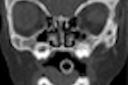
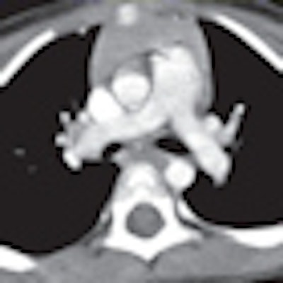
| Above images show a 3-year-old boy weighing 14.7 kg (32.3 lb) imaged with 120 kVp and measured noise of 9.6 HU. Images below are of a 4-year-old boy weighing 14.1 kg (31 lb) imaged with 80 kVp and measured noise of 11.3 HU. All transverse CT images with mediastinal and lung windows were rated as having good image quality. Images courtesy of the American Journal of Roentgenology. |
 |
The poor images were attributed to severe motion or catheter artifacts rather than related to changes in radiation dose. "All studies were considered diagnostically adequate," they wrote.
The researchers acknowledged several limitations of the study. They noted that study results could not be directly applied to different CT systems and different types of tube current modulation. In addition, the weight-based protocol had only two pediatric patient groups with the same reference mAs, "because we wanted a simple protocol that could be performed easily and reliably during routine chest CT."
They also wrote that the calculation of radiation dose is "fraught with problems and inaccuracies. The scanner-generated [CT dose index volume] and [dose-length product] were not specifically validated for this study, although the scanner undergoes routine checks and calibrations."
Even with the limitations, the researchers concluded that significant dose reduction could be achieved for routine pediatric chest CT "by paying attention to simple protocol adjustments. This is especially important for the younger, most radiation-sensitive children. The radiologists and clinicians accepted increased image noise as a trade-off for decreased dose, and no study was considered diagnostically compromised."
By Wayne Forrest
AuntMinnie.com staff writer
April 22, 2010
Related Reading
Automated exposure control delivers uneven CT dose reduction, April 15, 2010
CT doses, widely variable in Europe, are reduced by staff efforts, March 6, 2010
320-row CT minimizes dose in pediatric abdominal studies, March 3, 2010
CATCH rules accurately predict which kids need head CT, February 9, 2010
NIH tells scanner suppliers to include radiation monitoring, February 1, 2010
Copyright © 2010 AuntMinnie.com