The diagnostic capabilities of a dedicated extremity 1-tesla MRI scanner are comparable to those of higher-field-strength magnets when imaging menisci, ligaments, and other knee disorders, according to a study by New York researchers.
The study from New York University and the Hospital for Joint Diseases-Orthopaedic Institute in New York City also found that extremity MRI exceeds the accuracy of conventional MRI sequences at higher fields for cartilage, and may prove a cost-effective alternative to whole-body knee imaging.
Lead author Asheesh Harsha from New York University presented the results at the 2009 American Roentgen Ray Society (AARS) annual meeting in Boston.
In his presentation, Harsha noted that the majority of musculoskeletal MR imaging is conducted on closed 1.5-tesla systems, which have remained the gold standard for diagnosing internal derangements of the knee.
"Sensitivities and specificities have been reported between 80% and 100% for meniscal lesions and ACL [anterior cruciate ligament] tears," he added. "But MR exams using standard pulse sequences have not consistently been able to accurately grade and detect articular cartilage lesions. However, this is becoming more possible in MR imaging in higher fields."
The purpose of the study was to test the accuracy of a 1-tesla dedicated extremity MRI scanner for detecting, locating, and grading the extent of disorder to knee structures, including articular cartilage, menisci, and anterior cruciate ligaments.
The retrospective analysis reviewed MRI records of 99 patients who underwent MR exams of the knee (OrthOne, ONI Medical Systems, Wilmington, MA) between January 2005 and May 2008. All patients subsequently underwent knee arthroscopy by one of two board-certified orthopedic surgeons. In total, 616 exams were conducted through the 29-month study period on 68 men and 31 women, who had an average age of 52 years.
Surgical confirmation
All surgeries occurred within three months of the exams, with a median of 21 days. A report was generated to document the integrity of the menisci, all cartilage surfaces, and interior and posterior cruciate ligament, Harsha said. A group of seven musculoskeletal radiologists evaluated the integrity of the structures prior to surgery, and one radiologist crafted another report to include the study indication and prior images.
The sensitivity, specificity, accuracy, positive predictive values, and negative predictive values were reported for the detection, location, and severity of injury to these structures.
The researchers found that the "dedicated extremity MR produced excellent results with the medial meniscus, with lower but good values for the lateral meniscus and ACL," Harsha said.
|
||||||||||||||||||||||||||||||||||||||||||||||||||||||||||||
"With this data, we concluded that dedicated extremity MR is a powerful diagnostic tool for evaluating internal derangements of the knee," Harsha said. "There is excellent diagnostic performance for fully characterizing menisci, ACL, and patellofemoral cartilage with poor but comparable results to conventional MR for lateral and medial compartment cartilage."
 |
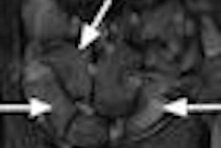
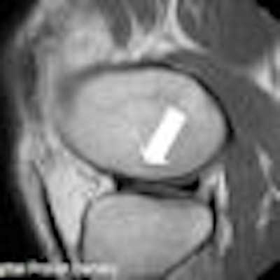
| Above, sagittal proton density-weighted image through the lateral meniscus from a 52-year-old male with interior knee pain. There is a short radial tear, which was seen only on the sagittal MR image and was present on the arthroscopy, making the image a true positive. Below, midsagittal proton density-weighted image is of the ACL of a 49-year-old male. The ACL appeared morphologically normal on MRI, but demonstrated laxity in arthroscopy. An orthopedic surgeon ruled a partial tear, making the image a false negative. Images courtesy of Asheesh Harsha and New York University. |
 |
Given the added benefits of improved patient comfort and safety, dedicated extremity MR "shows promise as a cost-effective addition or alternative to whole-body MR," he added. With dedicated extremity MR technology, the cost of siting and the cost of the magnet are both lower than with a conventional MR scanner.
By Wayne Forrest
AuntMinnie.com staff writer
June 24, 2009
Related Reading
Extremity 1T MRI equals 1.5T for rheumatoid arthritis, June 8, 2009
MRI with STIR fails to change diagnosis in child abuse cases, May 12, 2009
MRI helps find fractures among elderly female ED patients, April 29, 2009
MRI used to diagnose complex lung infections in children, October 16, 2008
MR parameters divulge depth of acute hamstring injuries, July 13, 2007
Copyright © 2009 AuntMinnie.com