Neurologists at Washington University School of Medicine in St. Louis are employing a functional MRI (fMRI) technique called resting state functional connectivity to predict the effects of strokes and other brain injuries on afflicted patients.
Resting state functional connectivity is designed to show the health of brain networks that are instrumental in allowing multiple parts of the brain to collaborate and function. The researchers say the technique might help determine the most appropriate treatment for stroke and brain injuries -- and later assess the effects of that treatment.
The study appears in the March issue of Annals of Neurology (2010, Vol. 67:3), with Dr. Alex Carter, Ph.D., assistant professor of neurology, as the lead author.
While resting state functional connectivity has been used for some 15 years, Carter said it has come into vogue more recently as an application to investigate a number of different disease states. The Washington University study is unique in its use of resting state functional connectivity to explore stroke and brain injuries.
Recovery clues
"From a clinical perspective, we still don't know all that much about what will determine how someone will recover from a traumatic brain injury or stroke, because there is a lot of variability in terms of how people do after their injuries," Carter said. "The idea was to use this type of imaging technique to look at brain networks to see if we could find clues that would help us do better in our prediction of who will do well or poorly, if we don't give them additional care and resources."
Researchers enrolled 23 first-time stroke patients (12 women and 11 men) with a mean age of 59.6 years. The study also included 11 healthy control subjects. All subjects were imaged on a 3-tesla MRI system (Magnetom Allegra, Siemens Healthcare, Malvern, PA).
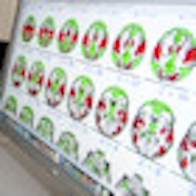
 |
| Dr. Alex Carter (left) and Dr. Maurizio Corbetta view MR images from their resting state functional connectivity technique to study brain organization and how it could help in treating brain injury patients. Image courtesy of Washington University School of Medicine. |
While traditional functional MRI requires the person in the scanner to perform a task to highlight the area of the brain under review, resting state functional connectivity only requires the patient to lie still and do nothing in order to obtain the desired brain images. In this study, patients were asked to simply relax and fixate on a central cross projected onto a screen at the head of the magnet bore.
"The reason that [having patients do nothing] is so crucially important is that it means we can study people who are too sick to follow very specific or complicated instructions," Carter said. "That is important if you want to study someone immediately after they have had a brain injury or a stroke."
Temporal correlation
In each patient, resting state functional connectivity measured the temporal correlation of the blood-oxygenation-level dependent (BOLD) signal across regions of the brain when not performing a task to determine the temporal coherence between brain regions. That information later was combined with the person's behavioral performance outside of the scanner to measure the performance of the brain networks under those conditions.
The researchers found that patients with damaged network connections between regions on different sides of the brain had greater functional impairments than patients with damaged connections between regions on the same side of the brain.
The MR images also confirmed that stroke damage on the left side of a patient's brain might cause control problems of his or her right arm, but the loss of coordination is significantly worse if the damage on the left side of the brain disrupted the network connections with the right side of the brain.
Right versus left
"Even though connectivity is lower in the left hemisphere that controls the right hand -- let's say, because you had a stroke in that hemisphere -- the connectivity that is most predictive of how that right hand will perform is the connectivity between the two hemispheres," Carter said. "We thought that was an interesting and novel finding that tells us something about how we need to approach people after they have had a brain injury or are recovering from a stroke."
He emphasized that this finding "does not contradict 100 years of research by any means. It simply says there are other layers that may be more subtle that we need to consider in terms of how the brain works."
Carter and his colleagues plan to continue their studies of brain injury and stroke patients and use resting state functional connectivity in long-term studies to see how well such patients recuperate.
"We would like to be able to use this technique to help us figure out who will do well and who will not do well in recovering from their stroke," he added. "For people who are at higher risk for a poor outcome, we can figure out what it is we need to give them, in terms of therapy, so they can have a more positive outcome."
The National Institute of Mental Health, the National Institute of Neurological Disorders and Stroke, the Robert Wood Johnson Foundation Amos Faculty Development Program, the James S. McDonnell Foundation, and the Rehabilitation Institute of St. Louis funded the research.
By Wayne Forrest
AuntMinnie.com staff writer
March 30, 2010
Related Reading
MRI shows impact of video games on brain regions, February 9, 2010
MRI shows that exercise may increase brain volume, February 1, 2010
Neuroimaging provides clearer picture of brain dysfunction in schizophrenia, August 5, 2009
fMRI helps find schizophrenia symptoms, January 20, 2009
MRI reveals patterns of neurological abnormalities in schizophrenia, April 4, 2006
Copyright © 2010 AuntMinnie.com